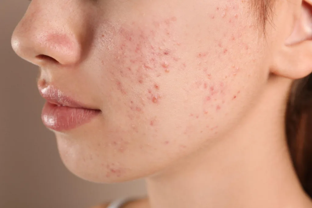
Key differences from acne and how we diagnose and treat it
Dr. Mara Padilla Evangelista-Huber, FPDS, FDSP, MClinRes
What are the main major differences between acne and “fungal acne”?
| Acne | Fungal acne | |
| Causes | Chronic inflammatory disorder of the hair follicles and the sebaceous glands.Occurs due to: Follicular hyper-keratinization (i.e. when old cells of the hair follicle do not shed normally onto the skin’s surface)Overproduction of oil /sebum (may be hormonally-related)Cutibacterium acnesInflammation | More appropriately called Malassezia folliculitis Malassezia – family of fungi that are often part of normal cutaneous flora. An overgrowth of this yeast can occur due to some factors, leading to skin conditions like tinea versicolor, folliculitis and seborrheic dermatitis. “Folliculitis” – inflammation of the hair follicle Although Malassezia folliculitis is caused by a fungus, it is not contagious. |
| Presentation | “Polymorphic” lesions Open and closed comedones, inflammatory papules (red bumps) and pustules (bumps with pus), and sometimes nodules and cysts | “Monomorphic” lesions Fine 1-2 millimeter papules and pustules in a follicular distribution (where the hair follicles are) |
| Location | Face, chest, shoulders, back | Face, chest, shoulders, back of arms and back (especially on areas of the body covered by occlusive clothing) If on face, upper forehead and hairline > central face |
| Other suggestive symptoms | Some variants get better with antibiotics targeting C. acnes Unaffected by antifungal therapy Not usually itchy | Persists or worsens despite the use of antibiotics targeting C. acnes (likely because these alter normal cutaneous flora, allowing for overgrowth of the fungus) Gets better with antifungal therapy Often itchy or burning |
Who is at risk of developing “fungal acne”?
- Adolescents (likely due to increased sebaceous gland activity)
- Individuals who are prone to increased sweating (e.g. patients with hyperhidrosis, athletes or those with frequent physical activity, overweight/obese individuals)
- Those with previous use of topical or oral antibiotics (e.g. tetracyclines) and oral corticosteroids and other immunosuppressive agents (e.g. chemotherapy, etc)
- Conditions with immunosuppression (e.g. AIDS, diabetes)
- Generally seems to be more common in males
Malassezia species are present on an estimated 92% of the world’s population as part of normal skin flora – but it does not overgrow in everyone who has it on their skin.
Can you distinguish “fungal acne” from acne with your naked eyes?
The diagnosis of Malassezia folliculitis is usually made based on the patient’s history and physical examination, but occasionally, there are challenging cases where further examination is warranted.
Because both acne and folliculitis affect the pilosebaceous unit, they can appear similar in presentation. In addition, like C. acnes, Malassezia has been shown to induce skin cells to generate inflammation via a similar pathway, adding to the potential overlap in clinical appearance.
What are the diagnostic tools commonly used to diagnose “fungal acne” and the treatment options?
Some of the diagnostic tools include:
- Scraping the skin lesion gently to get a sample of skin cells, and adding a substance called potassium hydroxide or KOH (KOH examination or smear) and looking at this under the microscope: positive findings are seeing abundant round, spores or budding yeast cells.
- Using a handheld device with black light (Wood’s light) to examine skin lesions – affected areas may fluoresce a bright yellow green color (some studies suggest blue-white color too) in a follicular pattern (where hair follicles are located).
- Taking a sample of your skin via a biopsy. The biopsy will show clusters of yeasts within the enlarged hair follicles surrounded by inflammatory cells. Staining the specimen with Periodic-Acid Schiff (PAS) stain may also aid in the visualization of the yeasts.
Treatment options:
- Topical antifungals (creams, body wash, shampoo) e.g. ketoconazole, clotrimazole, miconazole, ciclopirox olamine, etc
- Oral antifungals (fluconazole, itraconazole)
- Very limited evidence on isotretinoin
Because topical antifungals do not penetrate well into the hair follicle, first-line treatment is generally with oral antifungals. Improvement is expected within 1–2 months.
Supportive measures:
- Eliminate predisposing factors, if possible e.g. if the patient is on oral medications, discontinuation may result in improvement in the folliculitis without any specific treatment.
- Malassezia folliculitis may be chronic, and may wax and wane, depending on the weather, humidity, and the patient’s activity level.
- Wear loose clothing when it’s hot and humid.
- Change out of clothes immediately after working out or sweating a lot.
References:Rubenstein RM, Malerich SA. Malassezia (pityrosporum) folliculitis. J Clin Aesthet Dermatol. 2014;7(3):37-41.
Ayers K, Sweeney SM, Wiss K. Pityrosporum folliculitis: diagnosis and management in six female adolescents with acne vulgaris. Arch Pediatr Adolesc Med. 2005;159:64–67.
Gaitanis G, Velegraki A, Mayser P, et al. Skin diseases associated with Malassezia yeasts: facts and controversies. Clin Dermatol. 2013;31:455–463
Yu HJ, Lee SK, Son SJ, et al. Steroid acne vs Pityrosporum folliculitis: the incidence of Pityrosporum ovale and the effect of antifungal drugs in steroid acne. Int J Dermatol. 1998;37:772–777


