Value at your desk. Contact Us
Value at your desk. Contact Us
Carmela Augusta F. Dayrit-Castro, MD, FPDS
A “birthmark” is an irregularity on the skin which is visible at, or shortly after, birth. They are often benign and have no medical and psychological significance to the affected individual. However, there are some birthmarks for which medical attention should be sought. We name here a few of the more common types of birthmarks and mention when a child will need further evaluation by a physician.
BROWN BIRTHMARKS
Brown or pigmentary birthmarks are due to increased pigment or number of pigment producing cells in the skin.
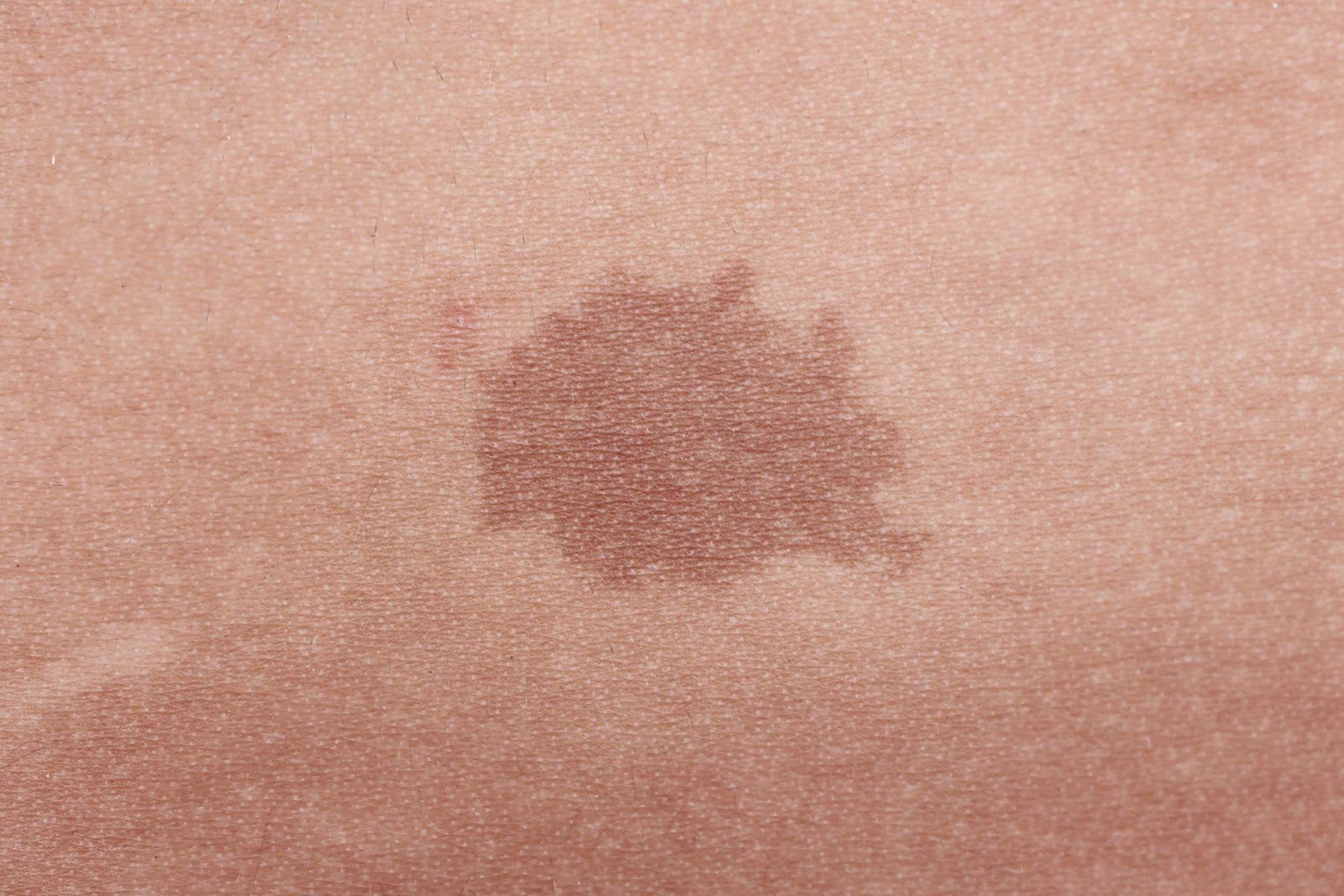
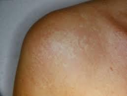
Café au lait macules (CALMs) are common and are seen as a solitary or a few, flat, light brown or tan spots on the skin. CALMs are benign but they tend to be permanent. If there are multiple CALMs found on the skin, further evaluation is necessary to assess for hereditary conditions like neurofibromatosis.
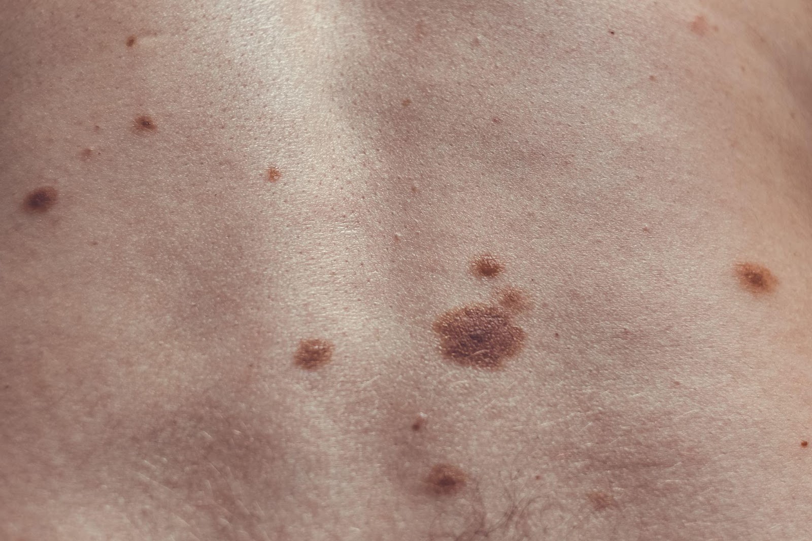
Congenital melanocytic nevi are typical moles which appear as discrete bumps or elevations which range in color from light to dark brown to black and may have overlying thin hairs. These moles vary in size from small to giant. They tend to increase in size proportionately as children grow. Children with large or giant congenital melanocytic nevi need to be evaluated since they may have an increased risk to develop melanoma and to have pigment cells in the nervous system.
BLUE BIRTHMARKS
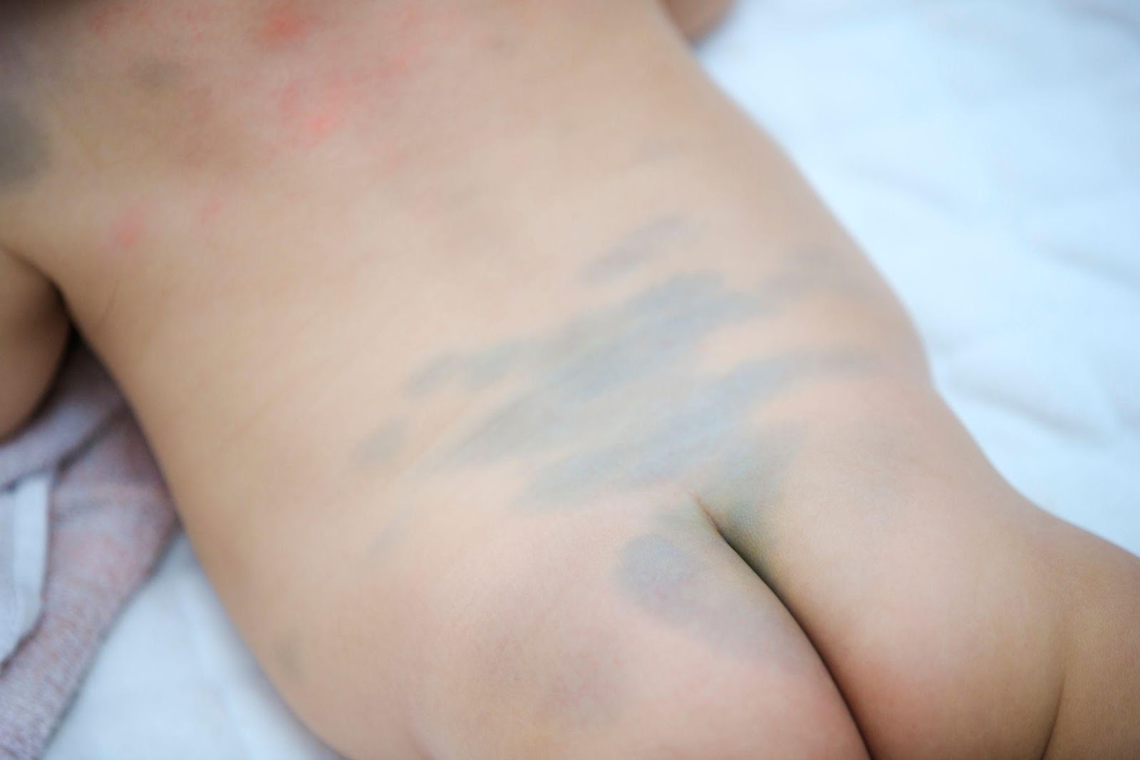
Dermal melanocytosis, commonly known as “Mongolian spots,” are blue-gray patches commonly located on the lower back and buttocks of infants. They appear more often in Asians and darker skinned individuals compared to lighter skinned individuals. These spots appear bluish because the pigment producing cells are halted on the lower layer of the skin, the dermis, en route to the upper layer, the epidermis. Mongolian spots tend to disappear by 3-5 years of age and rarely persist into adulthood. No further evaluation is needed for this. There are, however, pigment lasers that may be helpful for persistent dermal melanocytosis in adults.
RED BIRTHMARKS
Red birthmarks are due to the proliferation or growth of blood vessels on the skin.
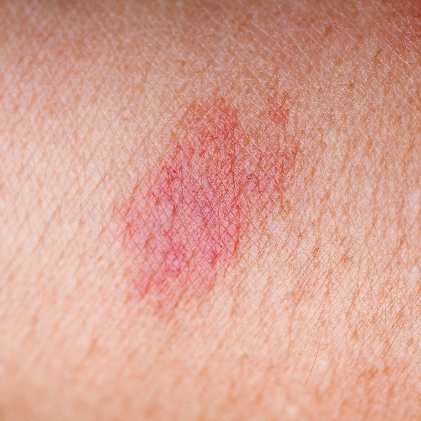
A nevus simplex, or commonly called “salmon patch,” “angel’s kiss,” or “stork bite” is a pink to red mark usually found between the eyebrows, forehead, upper eyelids and nape of newborns. They tend to become more visible when a baby is crying or after physical exertion. These are formed because of the dilation of the smallest blood vessels or capillaries. They tend to disappear between 1 to 3 years of age but the ones located on the nape tend to persist. No further evaluation is usually required for this.
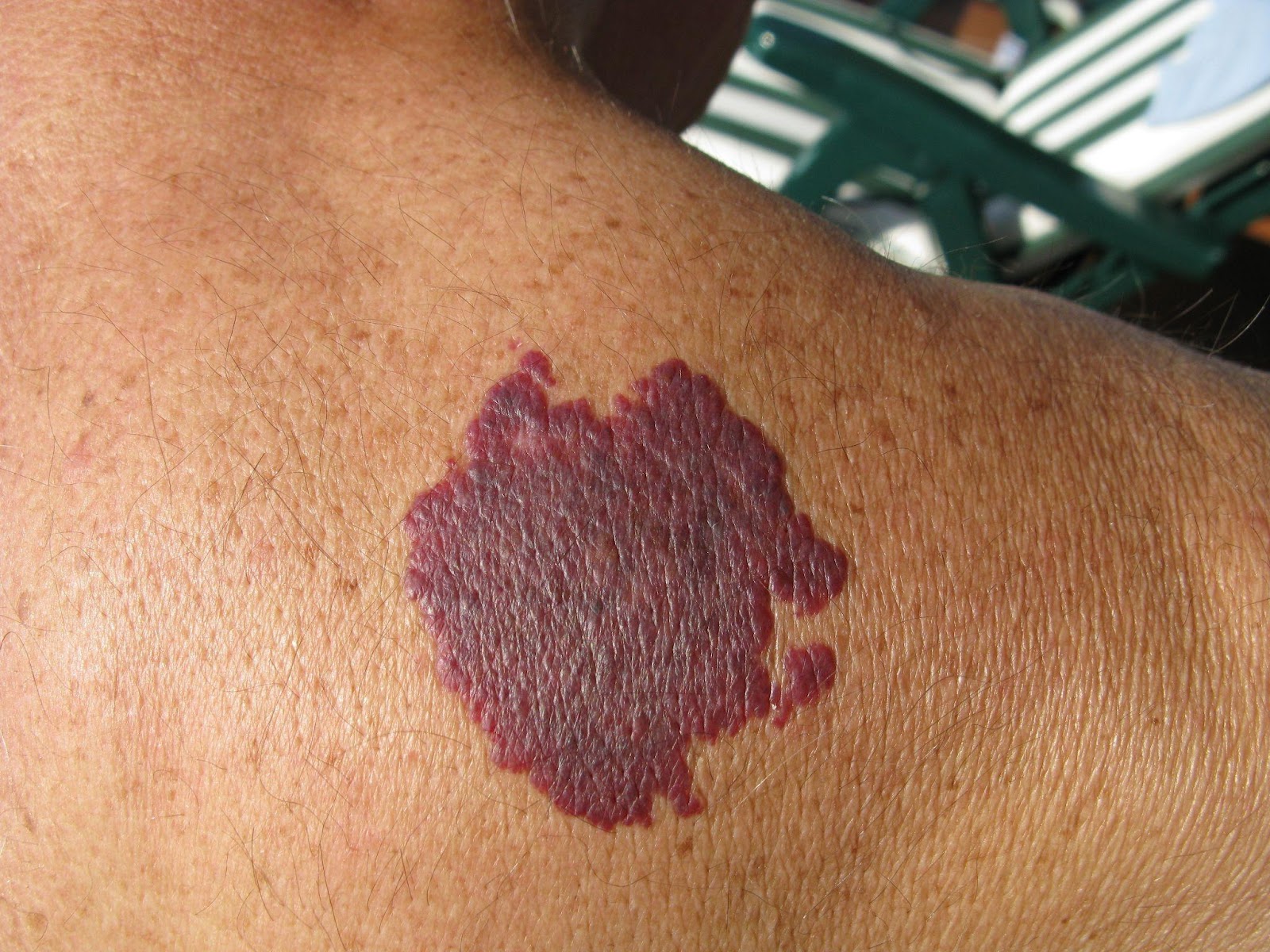
A port wine stain (PWS), is a more uniform, deeper red mark often located on the face. It can also appear elsewhere on the body, like on the arms and legs. A PWS appears because of an abnormal formation of capillaries on the skin, and may involve deeper tissues. Unlike a nevus simplex, PWSs tend to persist throughout life and may become thicker and develop red bumps on its surface during adolescence or adulthood. When a PWS appears on: (1) half of the face, (2) part of the face covering an upper or lower eyelid, (3) the midline of the forehead, or (4) a significant part of a limb, medical attention is recommended to assess involvement of the brain and eyes and discrepancy in limb size. There are also laser treatments available to help fade PWSs.
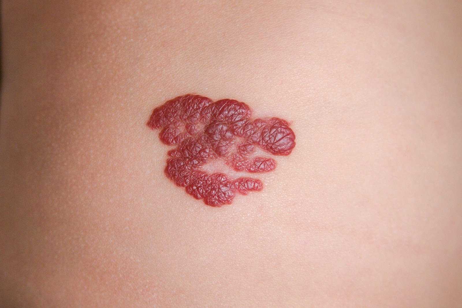
Infantile hemangiomas (IHs) often appear as soft, bright red bumps or elevations which are not, or barely, perceptible at birth. They are often mistaken as a mark from birth trauma. During the first 2 weeks of life, they begin to enlarge and may continue to enlarge until 3-5 months of age. Some IHs are deep and may appear as compressible skin colored or bluish green bumps that are recognized as infantile hemangiomas based on its growth pattern. Most IHs do not need treatment as they spontaneously resolve by 4-5 years of age. However, there are a subset of patients who need further evaluation and may require early treatment with oral medications during the first weeks of life. Early evaluation is needed when: (1) the IH is located on a “high-risk” location such as the eyelid, tip of nose, lip, midline chin and neck (beard area), ear, or groin as there is a risk for functional impairment, significant deformity, and ulceration; (2) there is a large IH on the face or on the lower back as this may be associated with other internal organ abnormalities; and (3) there are several infantile hemangiomas on the skin which could be a marker for having a liver hemangioma.
WHITE BIRTHMARKS
White birthmarks may appear due to decreased pigment or focal blood vessel constriction on the affected area.
Nevus depigmentosus is a whitish spot that is present since birth. They may be more difficult to see in lighter skinned individuals. There is decreased pigment or melanin in these spots. One spot is benign but if three or more are seen in a newborn, immediate evaluation for tuberous sclerosis complex should be sought.
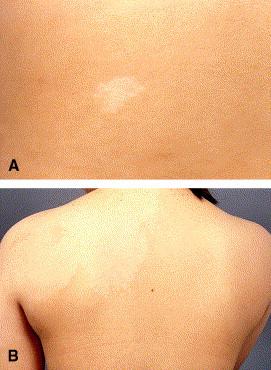
Nevus anemicus appears as a single or a group of whitish spots more often found on the trunk. They are asymptomatic and persist throughout life. It is an area of blood vessel constriction and when pressed at the border, can blend into the surrounding skin. As a solitary skin finding, no further evaluation is needed.
Other birthmarks
There may be less common and rare birthmarks which appear in various shapes, colors and textures—linear white or brown whorls and streaks; warty lesions; orange lesions on the scalp; tufts of hair; midline lesions. For more unusual birthmarks, your pediatrician may choose to refer you to a specialist for further evaluation and/or reassurance. But remember, when in doubt, it is always best to err on the side of caution and seek advice from your doctor.
REFERENCES:
Balin SJ. “Benign melanocytic neoplasms.” In: Bolognia JL, et al. Dermatology. (fourth edition). Mosby Elsevier, Spain, 2018:1954-88.
Baselga E. “Vascular malformations.” In: Bolognia JL, et al. Dermatology. (fourth edition). Mosby Elsevier, Spain, 2018:1805-27.
Chan YC. “Hypopigmentation disorders.” In: Eichenfield LF, et al. Neonatal and Infant Dermatology. (third edition). Mosby Elsevier, Spain, 2015: 369-87.
Hunt R “Neonatal dermatology.” In: Kang S, et al. Fitzpatrick’s Dermatology. (ninth edition). McGraw-Hill, USA, 2019: 1732-8.
Krowchuk DP, Frieden IJ, Mancini AJ, Darrow DH, Blei F, Greene AK, et al. Clinical Practice Guideline for the Management of Infantile Hemangiomas. Pediatrics. 2019 Jan;143(1):e20183475. doi: 10.1542/peds.2018-3475. PMID: 30584062.
Puttgen KB “Neonatal dermatology.” In: Cohen B. Pediatric Dermatology. (fourth edition). Elsevier, USA, 2013: 14-67.
by: Patricia Pontejos-Canivel, MD, DPDS
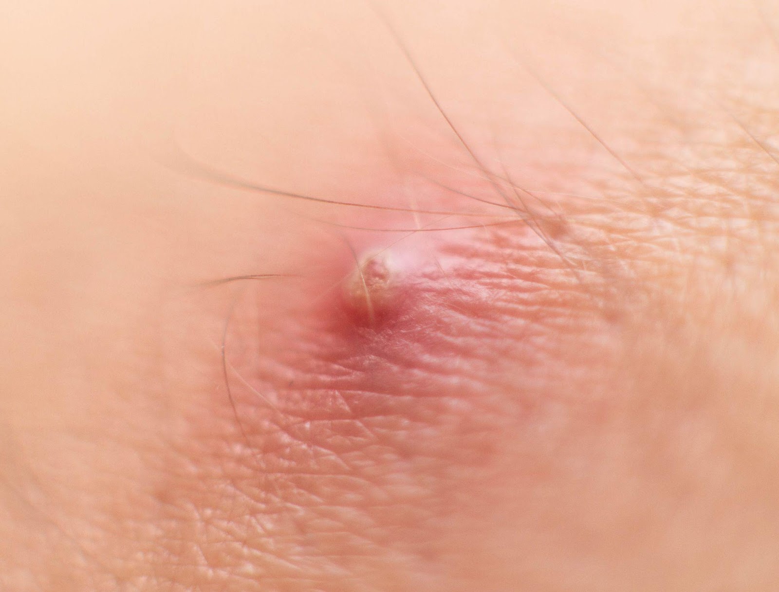
Boils are caused by a bacterial infection of the hair follicle. It presents as a painful red nodule on the skin that can start draining pus. This condition usually develops in areas that are prone to perspiration, friction and occlusion:
There are some factors that can predispose a person to developing boils such as:
Some boils may resolve on its own but if you are experiencing the following, consult a board- Dermatologist immediately:
Here are some tips to manage a boil at home:
References:
AAD website: https://www.aad.org/public/everyday-care/injured-skin/treat-boils-styes
Fitzpatrick’s Dermatology 9th edition
By Lonabel A. Encarnacion MD,FPDS, Elaine Marie Gutierrez-Villaroman MD,FPDS, Rizia Margate MD, Melissa See MD
Contact Dermatitis is skin inflammation or eczema that is triggered by substances that come in contact with the skin. These substances may be a chemical, biologic or a physical agent. Contact Dermatitis after a single or multiple exposure may be irritant or allergic. Let’s start with the more common irritant contact dermatitis (ICD) and later understand the more complex allergic contact dermatitis (ACD)
IRRITANT CONTACT DERMATITIS (ICD)
What is Irritant Contact Dermatitis?
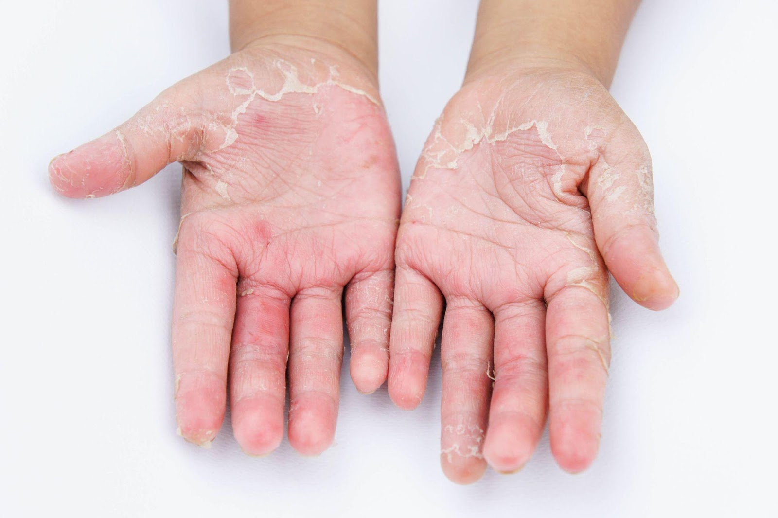
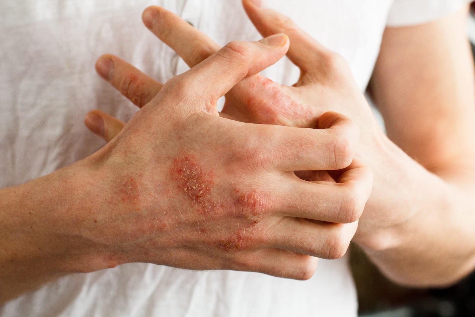
Irritant contact dermatitis or ICD is a nonspecific, nonallergic response of the skin to direct chemical damage. That means it’s a random, automatic skin response to a threat. ICD is a localized skin reaction to an irritant. Irritant chemicals are soaps, detergents, acids and alkalis, industrial solvents, even rough clothing, etc. It is the most common occupational skin disorder and hands are most often affected.
How do we get Irritant Contact Dermatitis?
ICD may be acquired from exposure to an irritant substance. This irritant is a corrosive agent that triggers release of inflammatory chemicals mainly from the upper layers of the skin. It is an immediate skin defense reaction.
Who can get affected with Irritant Contact Dermatitis?
Any individual who comes in contact with an irritant for a sufficient amount of time and adequate concentration will most likely have ICD.
For instance, an employed dishwasher whose occupation exposes him to constant soap & water work setting is prone to ICD. All of us now in this pandemic, who constantly wash hands, disinfect with alcohol or use bleach sanitizers are prone to ICD.
What are the usual signs & symptoms of Irritant Contact Dermatitis?
Clinical manifestations of ICD are stinging, burning and itching with reddish bumps. There could be redness, mild swelling and scaling. There could also be thickening & cracking of the skin with constant exposure with the irritant chemical.
How is Irritant Contact Dermatitis diagnosed?
Diagnosis of ICD is primarily clinical and rests on the exclusion of other cutaneous diseases especially allergic contact dermatitis. When a patch test is done and result is negative, that negative result is consistent with Irritant Contact Dermatitis. (Just read on!)
What is the treatment for Irritant Contact Dermatitis?
Protect the skin. Use Barrier creams. Use Personal Protective Equipment such as gloves.
If in contact with the corrosive chemical, right away wash-off with water. Topical/oral steroids are used. And on those itchy skin areas, massage with moisturizers instead of scratching. This calms that irritated & angry skin!
ALLERGIC CONTACT DERMATITIS (ACD)
What is Allergic Contact Dermatitis?
Allergic contact dermatitis or ACD is a “s-l-o-w” delayed type hypersensitivity reaction of the skin to an allergen. Contact allergens are like hair dye, metals, jewelry, rubber, topical medications, skin care products, plants, chemicals and many, many more. The skin may develop allergy to these environmental substances. It’s not immediate and it takes time.

How can we get Allergic Contact Dermatitis?
ACD may be acquired by contact to an allergen of a sensitized individual or someone who came in contact with the allergen days, weeks or even years prior to appearance of the lesions. This means a prior exposure to an allergen chemical initiates this skin sensitivity. It is not an immediate type of skin reaction.
Who can get affected with Allergic Contact Dermatitis?
Persons with persistent or relapsing dermatitis may have ACD. All ages can get affected with ACD but is usually uncommon in young children and seniors above 70 years old.
What are the usual signs & symptoms of Allergic Contact Dermatitis?
ACD is characterized by itchy reddish bumps and blisters sometimes oozing. Thickened itchy plaques indicate a longstanding condition. These lesions usually come and go.
How is Allergic Contact Dermatitis diagnosed?
Patch testing confirms ACD. This test involves the application of common allergens on the patient’s back to re-create and document skin reactivity. Remember, a positive patch test is able to identify the allergen which must definitely be avoided to resolve the disturbing, recurrent dermatitis.
What is the treatment of Allergic Contact Dermatitis?
Identify and remove the cause. This is the definitive management of this recurrent, disturbing skin condition known as Allergic Contact Dermatitis.
Topical and /or oral steroids are effective in controlling the signs and symptoms. And do consult a board-certified dermatologist for best advice to avoid the allergens!
References:
Medscape https://emedicine.medscape.com/article/1049216 updated Aug 20,2020. Allergic Contact Dermatitis Updated Aug 20, 2020 Author Thomas N Helm, MD
Medscape https://emedicine.medscape.com/article/1049353-overview
Irritant Contact Dermatitis Updated: Nov 20, 2020 Author: Savina Aneja, MD
Wolff K, Johnson RA, Saavedra A, Roh E. Fitzpatrick’s Color Atlas and Synopsis of Clinical Dermatology. 8th ed. New York: McGraw-Hill Medical. 2017: 20-33Kang S, Amagai M, et al. Fitzpatrick’s Dermatology. 9th ed. McGraw-Hill Education. 2019: 395-427
Pia Victoria Velasco, MD, FPDS
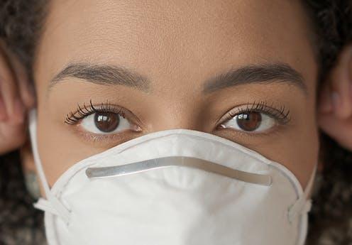
A. FACE MASK SKIN CARE – Masks play an important role in preventing the spread of Coronavirus. However, regular use of masks can cause skin problems such as acne, rashes and even itchiness. To prevent these skin problems, her are some helpful skin care tips:
Heavy make-up is more likely to clog your pores and lead to break outs. If you cannot skip your make-up, look for products labeled as “non-comedogenic” which will not clog your pores. Use make-up on the areas on the eye area only and skip it on the areas covered by the mask to lessen the risk of maskne.
B. HOW TO COPE WITH HAND DERMATITIS- Hand washing has been a mainstay in controlling the spread of Covid-19. Unfortunately, frequent hand washing and use of alcohol have often left hands feeling dry and irritated. Here are some helpful tips on how to keep Coronavirus away without compromising care for our hands:
References:
1. cdc.gov
2. pds.org: Recommendations for Addressing PPE-related Skin Care Issues during the COVID-19 Crisis
3. AAD COVID-19 Coronavirus Resource Center
Heirich Fevrier P. Manalili, RPh MD DPDS
Martha Joy Bruan-Tapales, RPh MD FPDS
There had been a lot of people (including doctors) who interchange a cream and an ointment. Knowing the difference between the two can help the patient and clinician decide on which preparation would benefit their condition more.
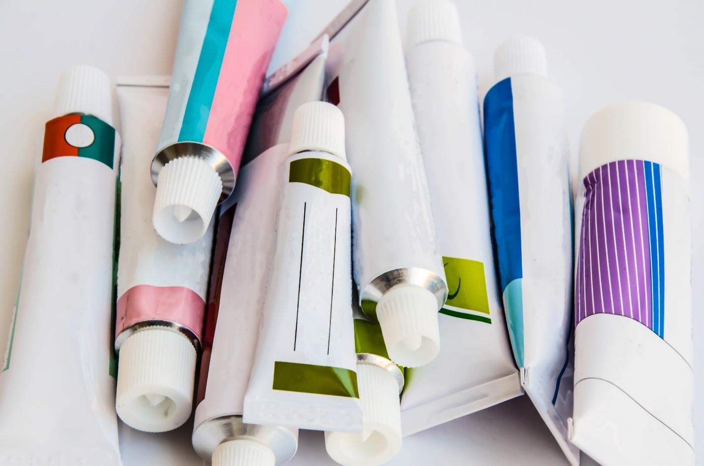
Ointments are semisolid preparations that contains lipid or hydrophobic ingredients intended for external application to the skin or other mucosal membranes1,2. It usually contains less than 20% of water and other volatile ingredients (eg. Ethanol), and more than 50% hydrocarbons and waxes1,2. They are designed to soften or melt at body temperature, spread easily, and have a smooth, non-gritty feel and appear translucent1. They are typically used as emollients to make skin pliable, barriers to prevent noxious substances from coming in contact to skin and vehicles for hydrophobic drugs1,2.
Creams are semisolid dosage forms containing one or more drug substances dissolved or dispersed in a suitable emulsion base1,2,3. They are more considered to be more fluid to other dosage forms1. They are usually found to have whitish, creamy appearance, due to scattering of light from dispersed phases (eg. Oil globules)1. Creams can either be on a water-in-oil emulsion (eg cold cream) which can be used as a softening and cleaning agent for make-ups1,2. On the other hand, it can also be in an oil-in-water emulsion (eg. Vanishing cream) which when rubbed on the skin, the water evaporates, leading to increased concentration of a water-soluble drug in the oily film which can adhere directly to the skin1,2.
Implications in Dermatology practice
As a vehicle, ointments have higher penetrability and are useful for thickened skin over palms and soles and over lichenified skin (eg. ichthyoses, psoriasis)4. The downside is that they are relatively greasy and messy to use. On the other hand, creams are less greasy and are more suited for moist and weeping areas of the skin (eg. Wounds with pus, blood and serum)4. Creams are preferred over ointments for mucosal areas because they are easier to spread and remove2.
In using topical steroids, the vehicle play an important role in determining the potency of the active ingredient4. For example, Mometasone furoate 0.1% cream is classified under Mid-potent (Class IV) while its counterpart Mometasone furoate 0.1% ointment is classified as high-potent (Class II).
References:
By: Bernadette Caluya, MD, DPDS
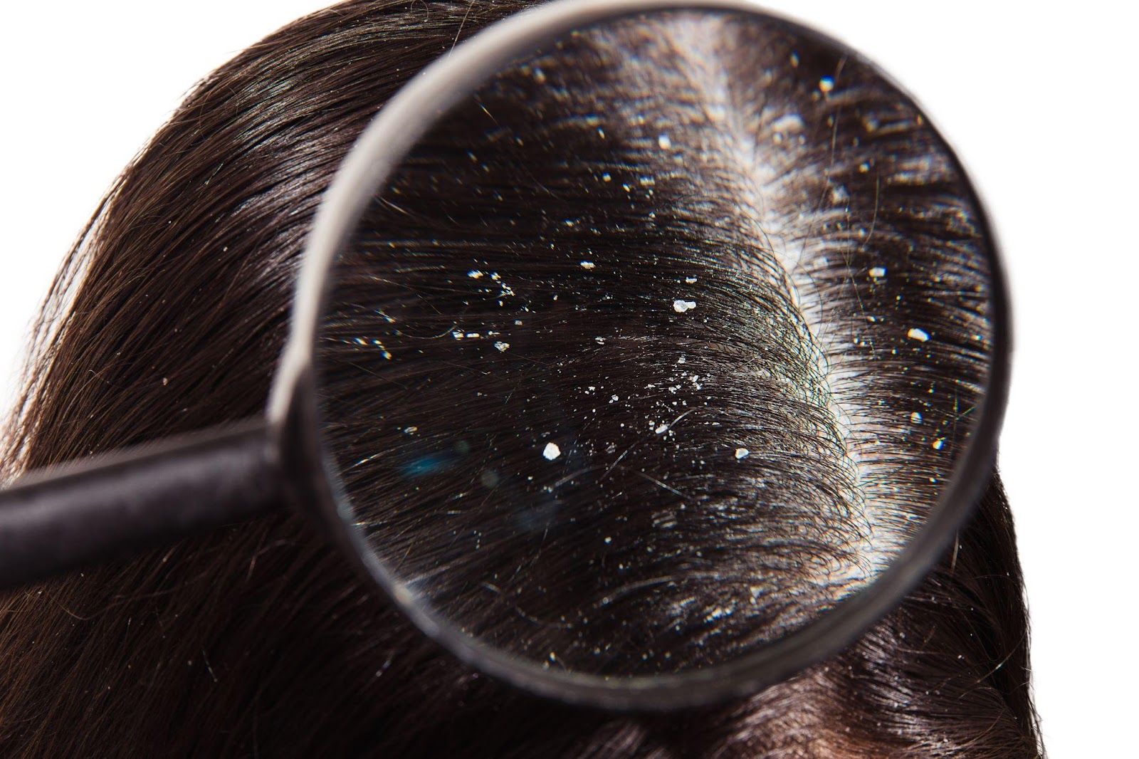
What is Dandruff?
Dandruff is a common skin condition where dry scales flake from the scalp. It is not contagious but it can be itchy and embarrassing to those who have it. It can occur across all age groups.It can be chronic and recurrent for many years. Dandruff may worsen during low temperature seasons and may also be associated with immunodeficiency and stress.
What are the symptoms?
It can be associated to the following symptoms:
-itchiness
-oily scalp
– scales on your scalp, eyebrows, back of ears, sides of the nose, chin, chest and upper back
What causes dandruff?
– seborrhea or increased sebum production in the scalp
– some cases are associated with increase of a fungus called Malassezia furfur on affected skin
– skin conditions like Seborrheic Dermatitis, Psoriasis, Eczema or Contact dermatitis to hair products
How to treat dandruff?
It is advisable to seek consult to a board-certified dermatologist before starting any treatment so that one’s condition will be assessed adequately and proper treatment will be prescribed.
Stephanie Katalbas-Asi, MD, FPDS
What is diaper rash?
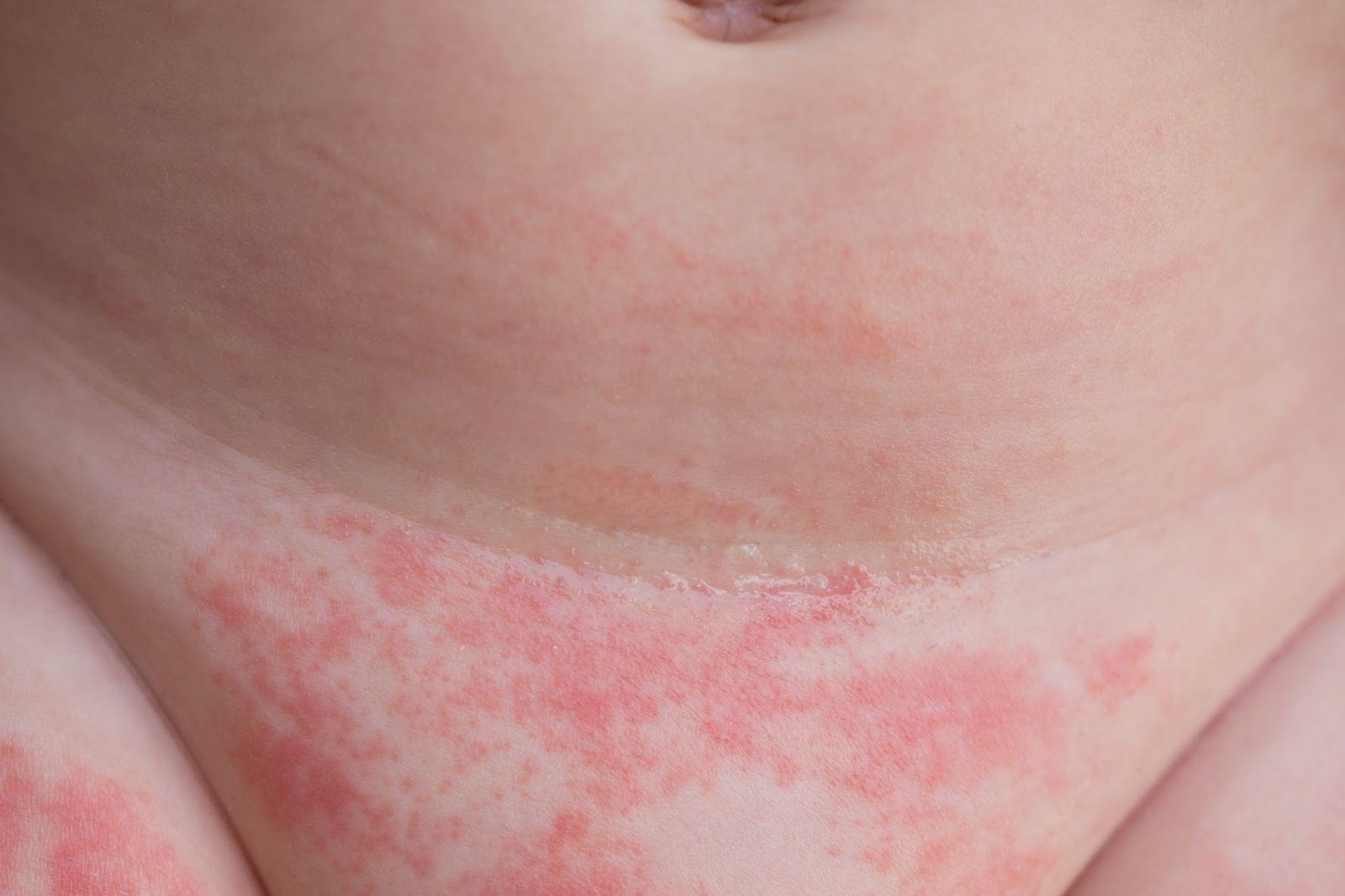
Diaper rash is a common form of reddish, inflamed skin on the areas covered by a diaper. This covers the genital area, buttocks, inner thighs, and sometimes the lower abdomen. This commonly affects babies, but it can also happen in adults who wear diapers.
In mild cases the skin can look pink, dry-looking skin, but in more severe cases it can resemble red, irritated, raw or burnt-looking skin with open wounds. It usually feels itchy or burning. Your child may seem more uncomfortable or irritable than usual, especially when changing diapers.
What causes diaper rash?
How is it treated?
The best thing to do is to keep the affected skin clean and dry as much as possible. Change diapers immediately after they are wet or soiled.
The exact treatment varies, depending on the exact cause for the diaper rash. This may include a mild steroid cream (e.g. hydrocortisone) and other antimicrobial creams. These medications should only be used under proper guidance from your doctor.
What can I do at home?
How to prevent diaper rash?
When should we see a doctor?
If the diaper rash doesn’t improve after 2-3 days with home treatment or shows any of the following changes, it’s best to consult a PDS certified Dermatologist for proper treatment.
Bibliography:
Stamatas, Georgios N., and Neena K. Tierney. “Diaper Dermatitis: Etiology, Manifestations, Prevention, and Management.” Pediatric Dermatology, vol. 31, no. 1, 2014, pp. 1-7.
by: Trixie Valle-Tin, MD, FPDS
What is Folliculitis?
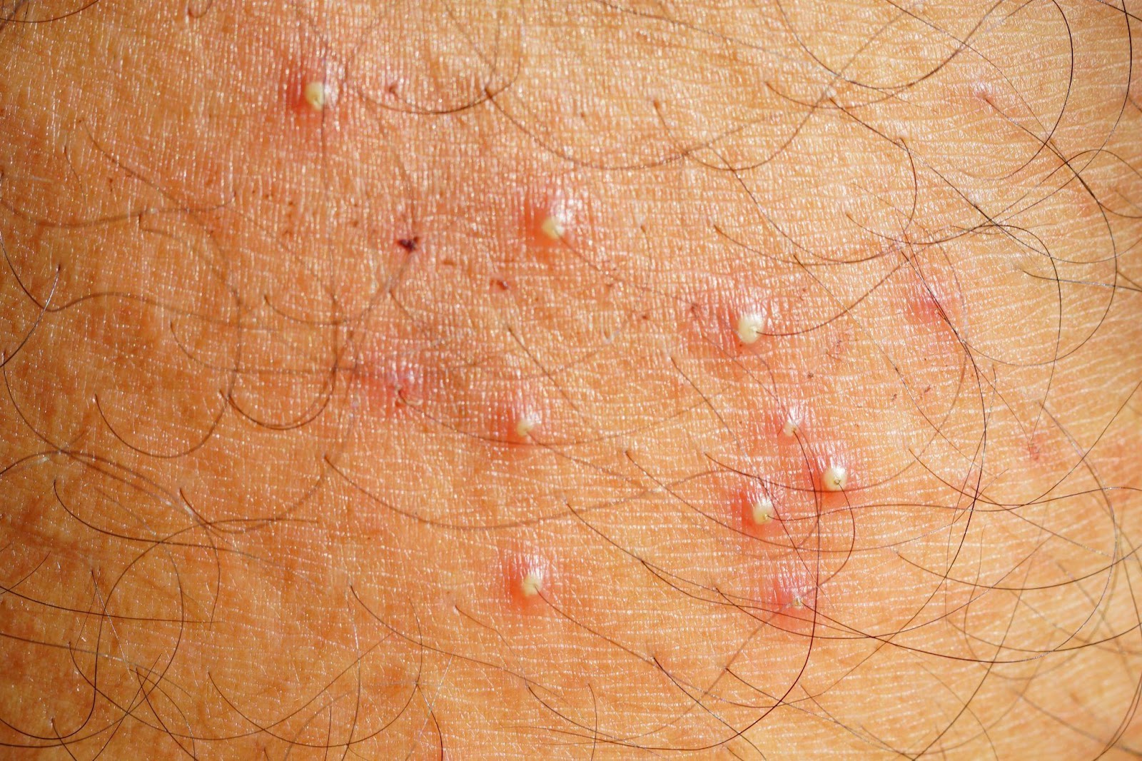
Folliculitis is an inflammatory process that involves the hair follicle or the pilosebaceous unit. It often manifests with redness, swelling, itching, or tenderness around the follicle and the perifollicular area. Folliculitis usually evolves in appearance and associated symptoms depending on the underlying cause and the depth of involvement. Clinically, it may appear as a dry thickened bump or as a pustule (pus-filled space) in the follicular opening or as a nodule deep in the follicle. It may also be a primary skin condition or as a secondary process in relation to another dermatologic disease. The location and the course of the folliculitis may help your dermatologist in determining the cause of the folliculitis.
What causes folliculitis?
Generally, folliculitis can either be infectious or non-infectious in origin.
Factors that can predispose to bacterial folliculitis include: conditions that lead to occlusion or maceration of the skin and exposure to contaminated surfaces, pre-existing itchy skin diseases that tend to be scratched such as eczema, and practices that may irritate the follicular unit such as improper hair removal. Nasal carriage of Staphylococcus aureus may also lead to repeated folliculitis in a person or his close contacts. Fungal folliculitis may occur with exposure to pets, soil, or another infected person, and usually appears in the setting of scaly red plaques with advancing borders that are typical of fungal infections. Folliculitis can also be seen in conditions of yeast overgrowth. Viral infections, most especially from the herpes virus, can also cause folliculitis and appear as pustular grouped skin lesions that tend to be recurrent in certain locations. A parasitic type of folliculitis may be caused by a mite such as Demodex that usually resides in the hair follicle and the sebaceous glands. When infection is a possible underlying cause of folliculitis, it is worthwhile to ask if the patient is immunosuppressed, or may be taking or applying immune-suppressing medications which may make the skin vulnerable to secondary infection if not taken under supervision of a doctor.
The non-infectious types of folliculitis are many and may be associated with genetics, gender, race and age and may be caused by several factors: stress, sun exposure, nutritional deficiency, hormones, drugs and medications, systemic illnesses such as diabetes or kidney disease, and occupational exposure to certain chemicals of which the most well-known are cutting oil, tar, DDT, and halogenated hydrocarbons. Chronic recurrent folliculitis may result from any of these factors or a combination thereof. Acne is a type of chronic folliculitis and the other conditions that may mimic acne like rosacea, perioral dermatitis, and acneiform eruptions have distinct clinical manifestations that must be differentiated from each other to be addressed appropriately.
How do we manage folliculitis?
The first step in managing folliculitis is to identify what causes it so it can be treated or avoided. Frequently, a thorough history and physical examination may be adequate to guide your board-certified dermatologist in giving the proper treatment. In some cases, certain diagnostic tests such as gram staining, culture, KOH smear, Tzanck smear, serologic tests, PCR, and histopathology may be warranted to arrive at the specific diagnosis.
When caused by infection, the treatment therefore is an antibacterial, antifungal, antiviral, or antiparasitic as deemed appropriate by a dermatologist. Any underlying skin disease such as an eczema or other conditions of impaired skin barrier must be treated as well. When the folliculitis is recurrent, it is important to identify external triggering factors such as occlusion, friction, chemicals, and improper shaving, but also internal and systemic factors such as nutrition, metabolic diseases, hormones, and immunosuppression among others. Regardless, consult with a board-certified dermatologist who can give the appropriate management of folliculitis.
How can we avoid getting folliculitis?
The following tips may help:
REFERENCES:
Luelmo-Aguilar, J., & Santandreu, M. S. (2004). Folliculitis: recognition and management. American journal of clinical dermatology, 5(5), 301–310. https://doi.org/10.2165/00128071-200405050-00003
Key differences from acne and how we diagnose and treat it
Dr. Mara Padilla Evangelista-Huber, FPDS, FDSP, MClinRes
What are the main major differences between acne and “fungal acne”?
| Acne | Fungal acne | |
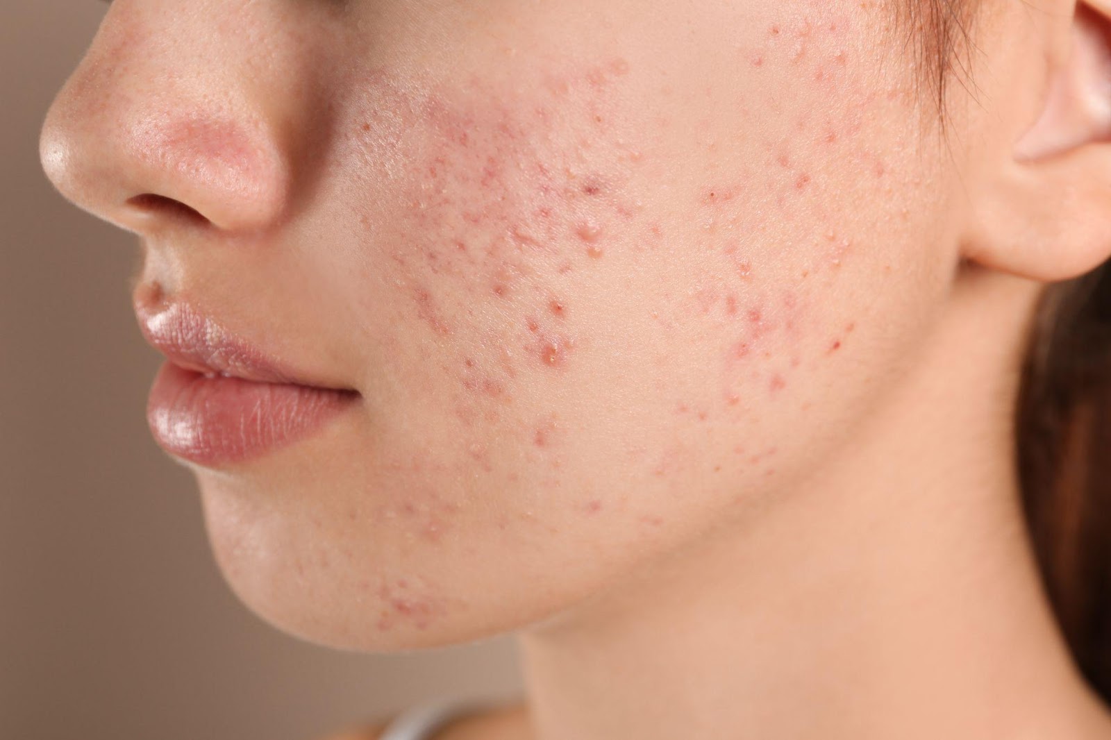
| Causes | Chronic inflammatory disorder of the hair follicles and the sebaceous glands.Occurs due to: Follicular hyper-keratinization (i.e. when old cells of the hair follicle do not shed normally onto the skin’s surface)Overproduction of oil /sebum (may be hormonally-related)Cutibacterium acnesInflammation | More appropriately called Malassezia folliculitis Malassezia – family of fungi that are often part of normal cutaneous flora. An overgrowth of this yeast can occur due to some factors, leading to skin conditions like tinea versicolor, folliculitis and seborrheic dermatitis. “Folliculitis” – inflammation of the hair follicle Although Malassezia folliculitis is caused by a fungus, it is not contagious. |
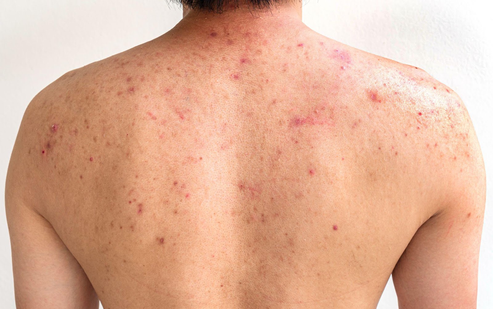
| Presentation | “Polymorphic” lesions Open and closed comedones, inflammatory papules (red bumps) and pustules (bumps with pus), and sometimes nodules and cysts | “Monomorphic” lesions Fine 1-2 millimeter papules and pustules in a follicular distribution (where the hair follicles are) |
| Location | Face, chest, shoulders, back | Face, chest, shoulders, back of arms and back (especially on areas of the body covered by occlusive clothing) If on face, upper forehead and hairline > central face |
| Other suggestive symptoms | Some variants get better with antibiotics targeting C. acnes Unaffected by antifungal therapy Not usually itchy | Persists or worsens despite the use of antibiotics targeting C. acnes (likely because these alter normal cutaneous flora, allowing for overgrowth of the fungus) Gets better with antifungal therapy Often itchy or burning |
Who is at risk of developing “fungal acne”?
Malassezia species are present on an estimated 92% of the world’s population as part of normal skin flora – but it does not overgrow in everyone who has it on their skin.
Can you distinguish “fungal acne” from acne with your naked eyes?
The diagnosis of Malassezia folliculitis is usually made based on the patient’s history and physical examination, but occasionally, there are challenging cases where further examination is warranted.
Because both acne and folliculitis affect the pilosebaceous unit, they can appear similar in presentation. In addition, like C. acnes, Malassezia has been shown to induce skin cells to generate inflammation via a similar pathway, adding to the potential overlap in clinical appearance.
What are the diagnostic tools commonly used to diagnose “fungal acne” and the treatment options?
Some of the diagnostic tools include:
Treatment options:
Because topical antifungals do not penetrate well into the hair follicle, first-line treatment is generally with oral antifungals. Improvement is expected within 1–2 months.
Supportive measures:
References:Rubenstein RM, Malerich SA. Malassezia (pityrosporum) folliculitis. J Clin Aesthet Dermatol. 2014;7(3):37-41.
Ayers K, Sweeney SM, Wiss K. Pityrosporum folliculitis: diagnosis and management in six female adolescents with acne vulgaris. Arch Pediatr Adolesc Med. 2005;159:64–67.
Gaitanis G, Velegraki A, Mayser P, et al. Skin diseases associated with Malassezia yeasts: facts and controversies. Clin Dermatol. 2013;31:455–463
Yu HJ, Lee SK, Son SJ, et al. Steroid acne vs Pityrosporum folliculitis: the incidence of Pityrosporum ovale and the effect of antifungal drugs in steroid acne. Int J Dermatol. 1998;37:772–777
Let’s talk About HPV, Baby!
By: Dr. Charina Ann R. Pelayo

Genital warts are the most common Sexually Transmitted Infections (STI) globally. They are caused by particular types of Human Papilloma Virus (HPV) and may be passed on through direct skin-to-skin contact with someone who has HPV on their skin during genital, oral or anal sex.
Genital warts may appear on the penis and scrotum in males, on the labia or in the vagina of females, and even on the anus and mouth or throat of both sexes. They may appear as flesh-colored bumps or look like small pieces of cauliflower. You can have just one genital wart or a bunch of them. Sometimes they itch, but most of the time you don’t feel anything at all. It is possible to get or even spread these warts even if you cannot see them. Although rare, a child may get infected with HPV while passing through the birth canal of the mother with genital warts.

Now, not all bumps on the genitals are caused by HPV. There are other skin conditions that might look like genital warts but are not. If you think you have genital warts, it’s important to get checked out by a board-certified dermatologist.

A dermatologist can diagnose genital warts by examining the warts during a consultation. Sometimes, the dermatologist will remove the genital wart or a part of it to send to the laboratory to confirm the diagnosis of HPV. For females, getting a PAP smear once a year is highly encouraged since HPV is the major cause for cancer of the cervix.
If you do get diagnosed with genital warts, there are several treatment modalities that can be done. A dermatologist may freeze or burn the warts in the clinic. He or she may also prescribe medicine that you will apply at home like Imiquimod, which increases the body’s immune system to get rid of the virus.
To protect yourself from getting infected with HPV, get vaccinated with the HPV vaccine. Practice safe sex. Latex condoms can lower your chances of getting HPV, but you may still get infected from the areas not covered by a condom.