Value at your desk. Contact Us
Value at your desk. Contact Us
Stephanie Katalbas-Asi, MD, FPDS
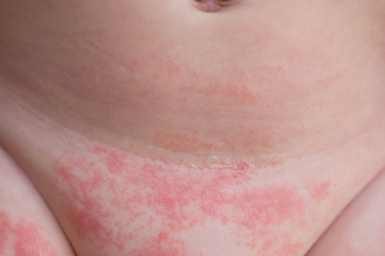
What is diaper rash?
Diaper rash is a common form of reddish, inflamed skin on the areas covered by a diaper. This covers the genital area, buttocks, inner thighs, and sometimes the lower abdomen. This commonly affects babies, but it can also happen in adults who wear diapers.
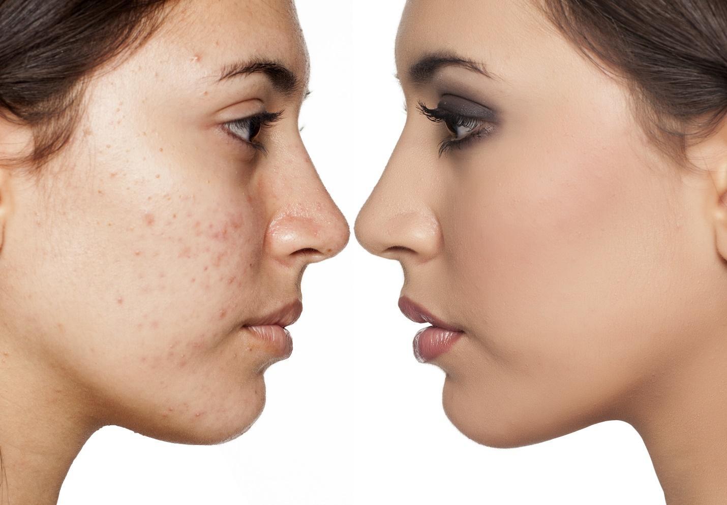
In mild cases the skin can look pink, dry-looking skin, but in more severe cases it can resemble red, irritated, raw or burnt-looking skin with open wounds. It usually feels itchy or burning. Your child may seem more uncomfortable or irritable than usual, especially when changing diapers.
What causes diaper rash?
How is it treated?
The best thing to do is to keep the affected skin clean and dry as much as possible. Change diapers immediately after they are wet or soiled.
The exact treatment varies, depending on the exact cause for the diaper rash. This may include a mild steroid cream (e.g. hydrocortisone) and other antimicrobial creams. These medications should only be used under proper guidance from your doctor.
What can I do at home?
How to prevent diaper rash?
When should we see a doctor?
If the diaper rash doesn’t improve after 2-3 days with home treatment or shows any of the following changes, it’s best to consult a PDS certified Dermatologist for proper treatment.
Bibliography:
Stamatas, Georgios N., and Neena K. Tierney. “Diaper Dermatitis: Etiology, Manifestations, Prevention, and Management.” Pediatric Dermatology, vol. 31, no. 1, 2014, pp. 1-7.
by: Trixie Valle-Tin, MD, FPDS
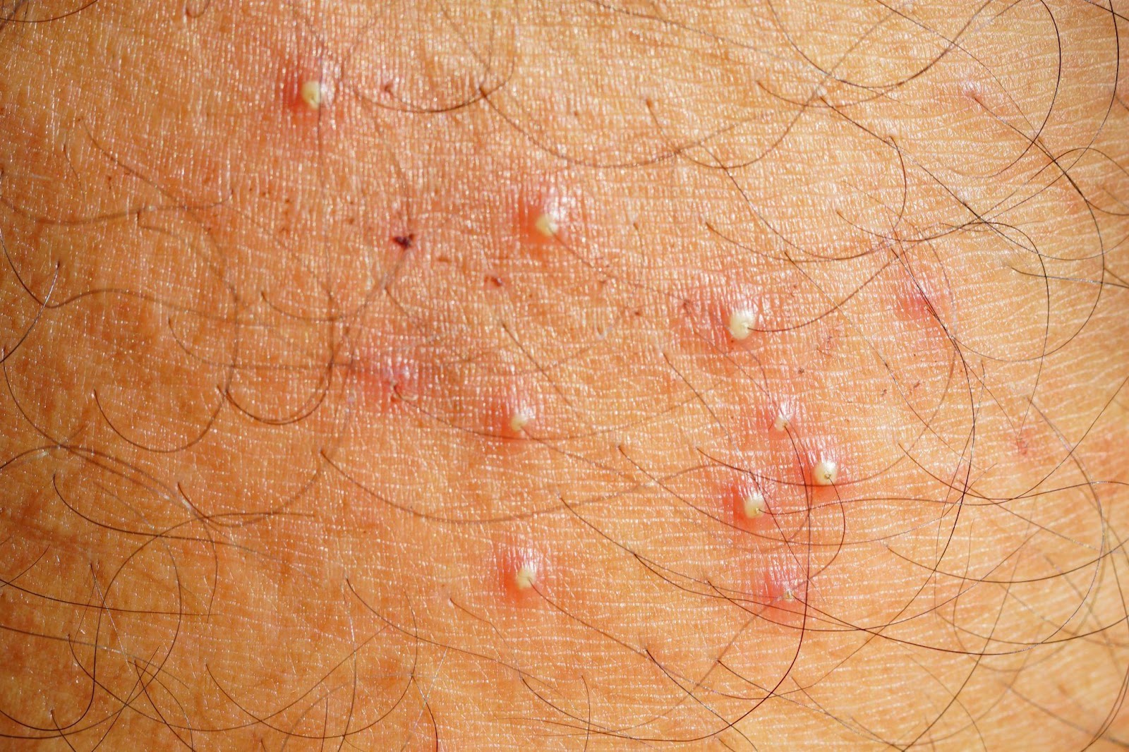
What is Folliculitis?
Folliculitis is an inflammatory process that involves the hair follicle or the pilosebaceous unit. It often manifests with redness, swelling, itching, or tenderness around the follicle and the perifollicular area. Folliculitis usually evolves in appearance and associated symptoms depending on the underlying cause and the depth of involvement. Clinically, it may appear as a dry thickened bump or as a pustule (pus-filled space) in the follicular opening or as a nodule deep in the follicle. It may also be a primary skin condition or as a secondary process in relation to another dermatologic disease. The location and the course of the folliculitis may help your dermatologist in determining the cause of the folliculitis.
What causes folliculitis?
Generally, folliculitis can either be infectious or non-infectious in origin.
Factors that can predispose to bacterial folliculitis include: conditions that lead to occlusion or maceration of the skin and exposure to contaminated surfaces, pre-existing itchy skin diseases that tend to be scratched such as eczema, and practices that may irritate the follicular unit such as improper hair removal. Nasal carriage of Staphylococcus aureus may also lead to repeated folliculitis in a person or his close contacts. Fungal folliculitis may occur with exposure to pets, soil, or another infected person, and usually appears in the setting of scaly red plaques with advancing borders that are typical of fungal infections. Folliculitis can also be seen in conditions of yeast overgrowth. Viral infections, most especially from the herpes virus, can also cause folliculitis and appear as pustular grouped skin lesions that tend to be recurrent in certain locations. A parasitic type of folliculitis may be caused by a mite such as Demodex that usually resides in the hair follicle and the sebaceous glands. When infection is a possible underlying cause of folliculitis, it is worthwhile to ask if the patient is immunosuppressed, or may be taking or applying immune-suppressing medications which may make the skin vulnerable to secondary infection if not taken under supervision of a doctor.
The non-infectious types of folliculitis are many and may be associated with genetics, gender, race and age and may be caused by several factors: stress, sun exposure, nutritional deficiency, hormones, drugs and medications, systemic illnesses such as diabetes or kidney disease, and occupational exposure to certain chemicals of which the most well-known are cutting oil, tar, DDT, and halogenated hydrocarbons. Chronic recurrent folliculitis may result from any of these factors or a combination thereof. Acne is a type of chronic folliculitis and the other conditions that may mimic acne like rosacea, perioral dermatitis, and acneiform eruptions have distinct clinical manifestations that must be differentiated from each other to be addressed appropriately.
How do we manage folliculitis?
The first step in managing folliculitis is to identify what causes it so it can be treated or avoided. Frequently, a thorough history and physical examination may be adequate to guide your board-certified dermatologist in giving the proper treatment. In some cases, certain diagnostic tests such as gram staining, culture, KOH smear, Tzanck smear, serologic tests, PCR, and histopathology may be warranted to arrive at the specific diagnosis.
When caused by infection, the treatment therefore is an antibacterial, antifungal, antiviral, or antiparasitic as deemed appropriate by a dermatologist. Any underlying skin disease such as an eczema or other conditions of impaired skin barrier must be treated as well. When the folliculitis is recurrent, it is important to identify external triggering factors such as occlusion, friction, chemicals, and improper shaving, but also internal and systemic factors such as nutrition, metabolic diseases, hormones, and immunosuppression among others. Regardless, consult with a board-certified dermatologist who can give the appropriate management of folliculitis.
How can we avoid getting folliculitis?
The following tips may help:
REFERENCES:
Luelmo-Aguilar, J., & Santandreu, M. S. (2004). Folliculitis: recognition and management. American journal of clinical dermatology, 5(5), 301–310. https://doi.org/10.2165/00128071-200405050-00003
Key differences from acne and how we diagnose and treat it
Dr. Mara Padilla Evangelista-Huber, FPDS, FDSP, MClinRes
What are the main major differences between acne and “fungal acne”?
| Acne | Fungal acne | |
| Causes | Chronic inflammatory disorder of the hair follicles and the sebaceous glands.Occurs due to: Follicular hyper-keratinization (i.e. when old cells of the hair follicle do not shed normally onto the skin’s surface)Overproduction of oil /sebum (may be hormonally-related)Cutibacterium acnesInflammation | More appropriately called Malassezia folliculitis Malassezia – family of fungi that are often part of normal cutaneous flora. An overgrowth of this yeast can occur due to some factors, leading to skin conditions like tinea versicolor, folliculitis and seborrheic dermatitis. “Folliculitis” – inflammation of the hair follicle Although Malassezia folliculitis is caused by a fungus, it is not contagious. |
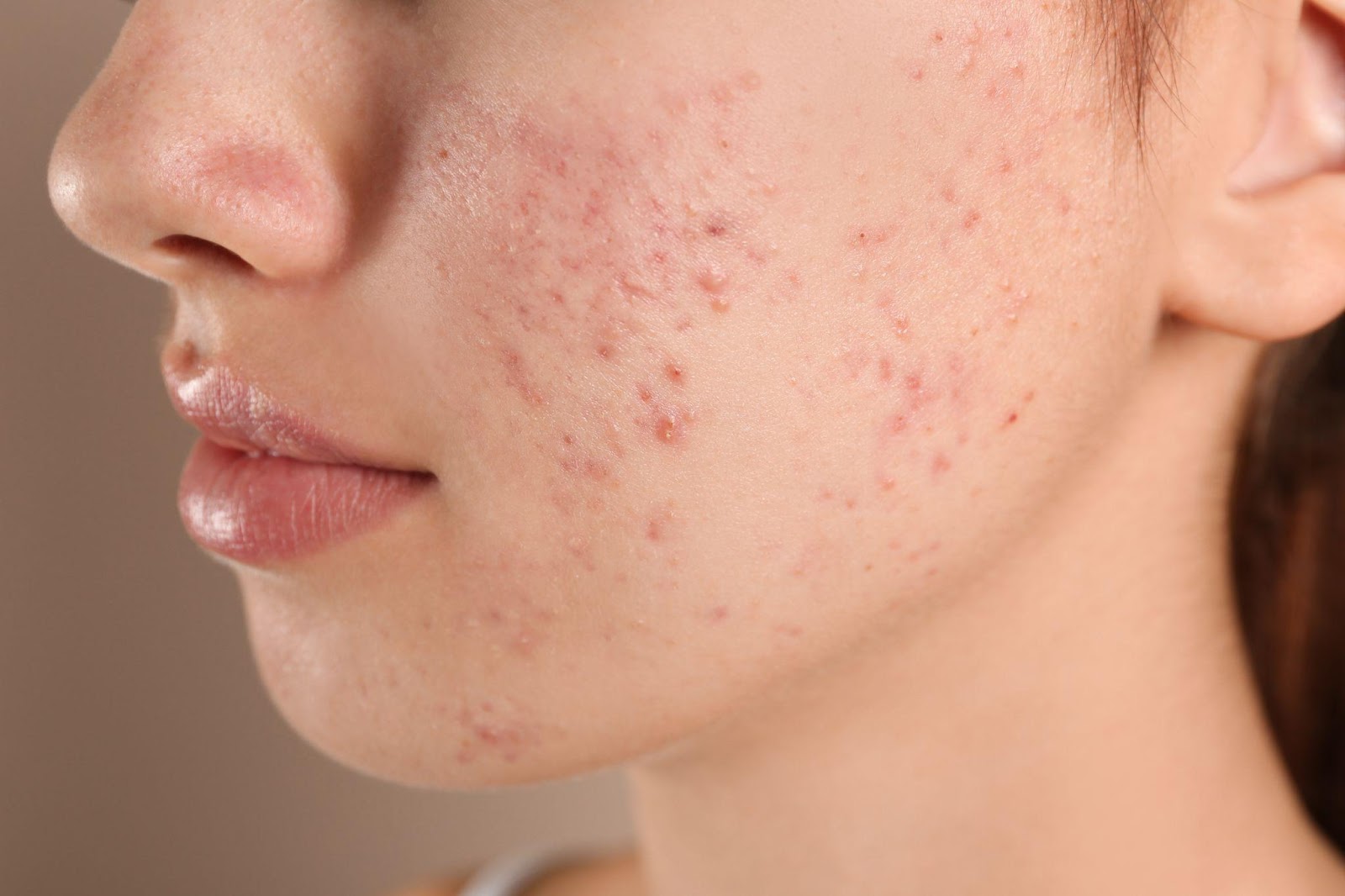
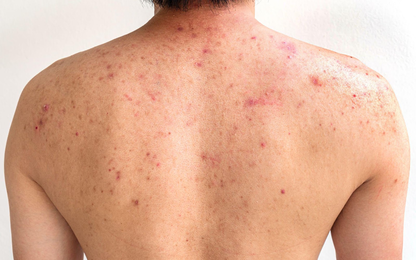
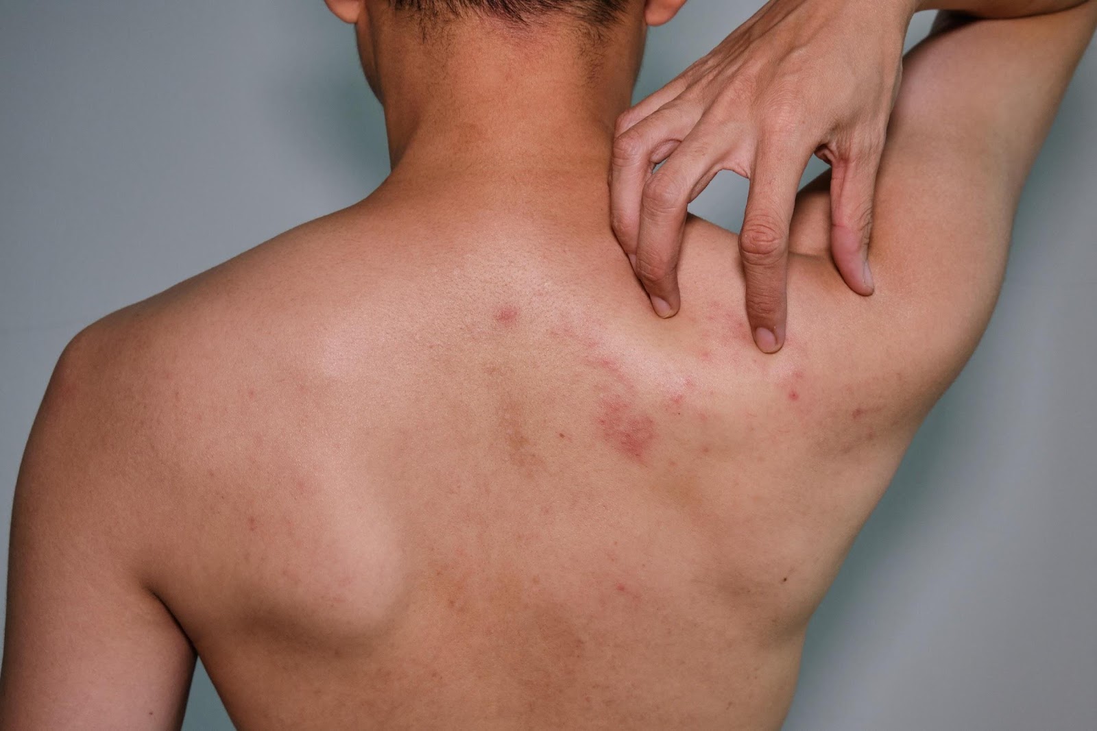
| Presentation | “Polymorphic” lesions Open and closed comedones, inflammatory papules (red bumps) and pustules (bumps with pus), and sometimes nodules and cysts | “Monomorphic” lesions Fine 1-2 millimeter papules and pustules in a follicular distribution (where the hair follicles are) |
| Location | Face, chest, shoulders, back | Face, chest, shoulders, back of arms and back (especially on areas of the body covered by occlusive clothing) If on face, upper forehead and hairline > central face |
| Other suggestive symptoms | Some variants get better with antibiotics targeting C. acnes Unaffected by antifungal therapy Not usually itchy | Persists or worsens despite the use of antibiotics targeting C. acnes (likely because these alter normal cutaneous flora, allowing for overgrowth of the fungus) Gets better with antifungal therapy Often itchy or burning |
Who is at risk of developing “fungal acne”?
Malassezia species are present on an estimated 92% of the world’s population as part of normal skin flora – but it does not overgrow in everyone who has it on their skin.
Can you distinguish “fungal acne” from acne with your naked eyes?
The diagnosis of Malassezia folliculitis is usually made based on the patient’s history and physical examination, but occasionally, there are challenging cases where further examination is warranted.
Because both acne and folliculitis affect the pilosebaceous unit, they can appear similar in presentation. In addition, like C. acnes, Malassezia has been shown to induce skin cells to generate inflammation via a similar pathway, adding to the potential overlap in clinical appearance.
What are the diagnostic tools commonly used to diagnose “fungal acne” and the treatment options?
Some of the diagnostic tools include:
Treatment options:
Because topical antifungals do not penetrate well into the hair follicle, first-line treatment is generally with oral antifungals. Improvement is expected within 1–2 months.
Supportive measures:
References:Rubenstein RM, Malerich SA. Malassezia (pityrosporum) folliculitis. J Clin Aesthet Dermatol. 2014;7(3):37-41.
Ayers K, Sweeney SM, Wiss K. Pityrosporum folliculitis: diagnosis and management in six female adolescents with acne vulgaris. Arch Pediatr Adolesc Med. 2005;159:64–67.
Gaitanis G, Velegraki A, Mayser P, et al. Skin diseases associated with Malassezia yeasts: facts and controversies. Clin Dermatol. 2013;31:455–463
Yu HJ, Lee SK, Son SJ, et al. Steroid acne vs Pityrosporum folliculitis: the incidence of Pityrosporum ovale and the effect of antifungal drugs in steroid acne. Int J Dermatol. 1998;37:772–777
Let’s talk About HPV, Baby!
By: Dr. Charina Ann R. Pelayo
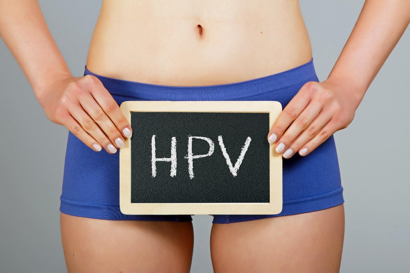
Genital warts are the most common Sexually Transmitted Infections (STI) globally. They are caused by particular types of Human Papilloma Virus (HPV) and may be passed on through direct skin-to-skin contact with someone who has HPV on their skin during genital, oral or anal sex.
Genital warts may appear on the penis and scrotum in males, on the labia or in the vagina of females, and even on the anus and mouth or throat of both sexes. They may appear as flesh-colored bumps or look like small pieces of cauliflower. You can have just one genital wart or a bunch of them. Sometimes they itch, but most of the time you don’t feel anything at all. It is possible to get or even spread these warts even if you cannot see them. Although rare, a child may get infected with HPV while passing through the birth canal of the mother with genital warts.

Now, not all bumps on the genitals are caused by HPV. There are other skin conditions that might look like genital warts but are not. If you think you have genital warts, it’s important to get checked out by a board-certified dermatologist.

A dermatologist can diagnose genital warts by examining the warts during a consultation. Sometimes, the dermatologist will remove the genital wart or a part of it to send to the laboratory to confirm the diagnosis of HPV. For females, getting a PAP smear once a year is highly encouraged since HPV is the major cause for cancer of the cervix.
If you do get diagnosed with genital warts, there are several treatment modalities that can be done. A dermatologist may freeze or burn the warts in the clinic. He or she may also prescribe medicine that you will apply at home like Imiquimod, which increases the body’s immune system to get rid of the virus.
To protect yourself from getting infected with HPV, get vaccinated with the HPV vaccine. Practice safe sex. Latex condoms can lower your chances of getting HPV, but you may still get infected from the areas not covered by a condom.
Corazon Almira Mella, MD, DPDS
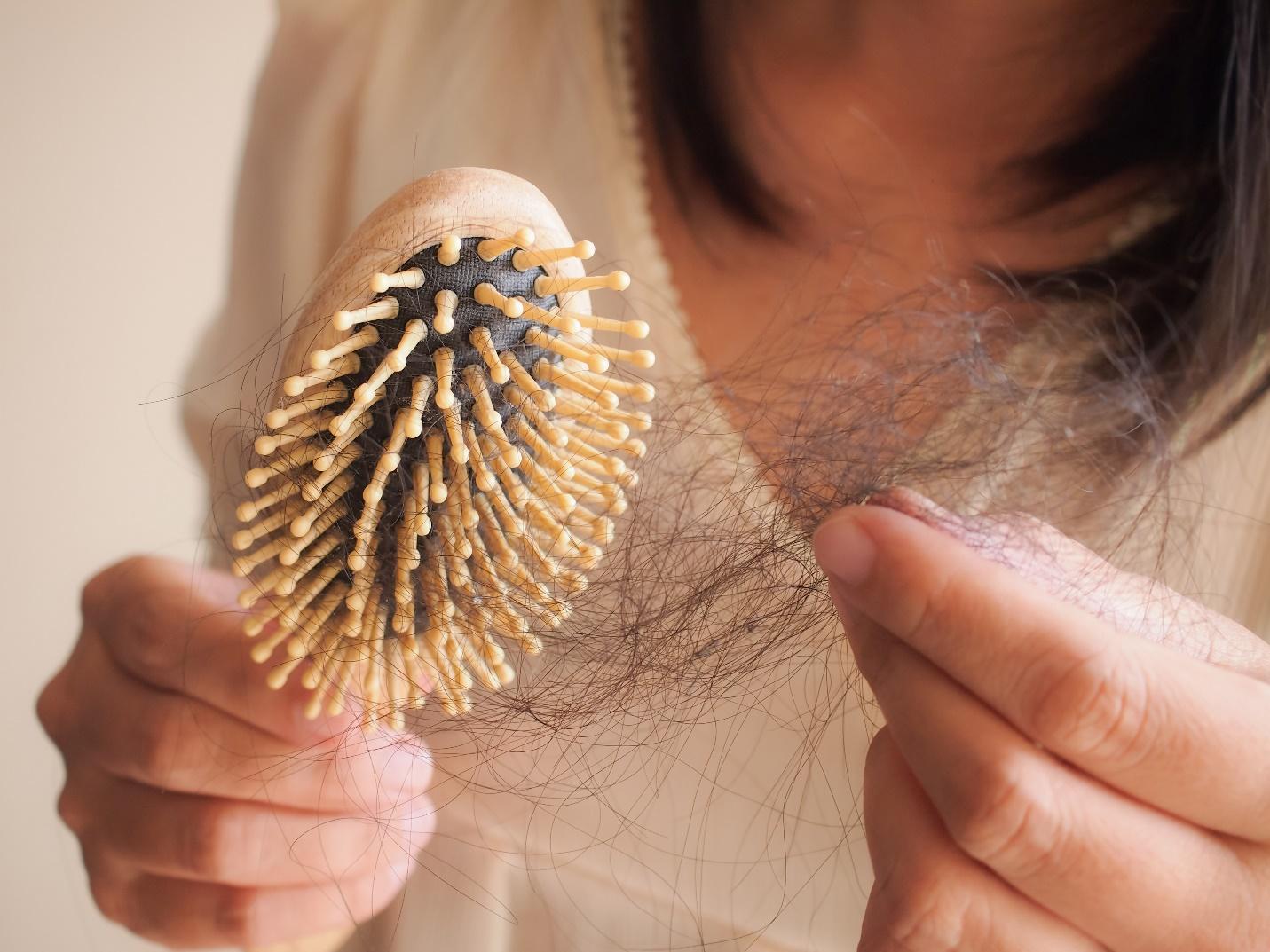
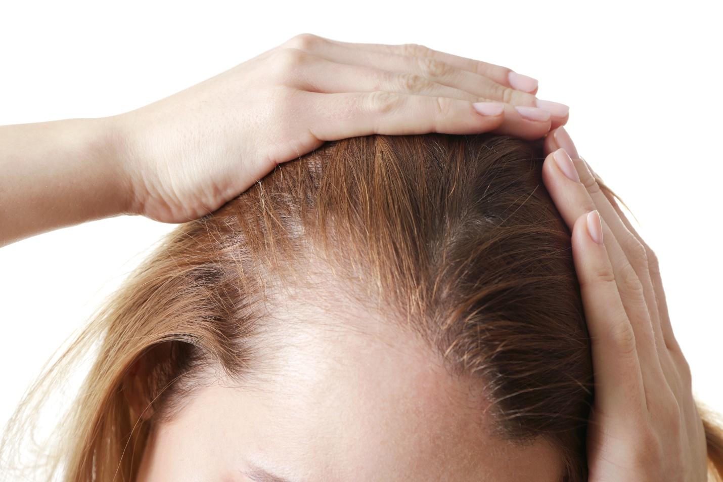
Hair loss among women remains a “taboo” topic. Unlike with men wherein it is considered socially acceptable with advancing age, women carry the pressure of maintaining a full scalp of hair throughout life. The notion that hair is associated with one’s beauty, as old-fashioned as it seems, still encompasses today’s culture. But just like men, women also suffer from hair thinning and hair loss – medically termed as female pattern hair loss (FPHL). And, it’s about time the discussion on such topic be normalized – because the more women are aware of it, the more that they can take control. Moreover, the earlier the hair loss is acknowledged and addressed, the better the outcome will be.
What is female pattern hair loss?
FPHL is the most common cause of hair loss among women. It can affect any age group but is more widely seen after menopause. It is characterized by progressive and widespread thinning of hair over the top of the head or crown. Dermatologists refer to this as the “Christmas tree” pattern but the younger generation have termed it is as hair or scalp cleavage. In contrast with male hair loss, female pattern hair loss does not usually result in total baldness. The process usually occurs in bouts – wherein there are accelerated periods of hair loss of around 3-6 months that are followed by episodes of stability that can last a year.
What causes female pattern hair loss?
Now, there are many reasons for female hair loss and this not only includes FPHL but also other medical conditions, physical and emotional stress. To be able to rule out other possible causes, a consult with a board certified dermatologist should be done.
The condition has a strong genetic predisposition and these genes could be inherited from either or both parents. Unlike with male hair loss, it is not clear whether or not androgens or male sex hormones play a role in its development. Environmental factors such as psychological stress, hypertension, diabetes mellitus, smoking, lack of photoprotection and physical activity have also been noted to be possibly related to FPHL.
Are there diagnostic tests that needs to be done for female pattern hair loss?
To properly investigate the root cause of hair loss, several tests are done by the dermatologist. This may include a hair pull test which is done by gently pulling one’s hair to evaluate how many hairs come out, blood examinations to check for vitamin, mineral and hormone levels as well as scalp examination and trichoscopy to rule out other causes of hair loss. In select cases, a scalp biopsy might also be done by a dermatologist. Biopsies are done to exclude more other types of hair loss such as scarring alopecias (scarring alopecias leads to permanent hair loss).
What is the treatment for female pattern hair loss?
Although there is no absolute cure for female pattern hair loss, there are several treatments available. These treatments are mainly done to slow down or stop the progression of hair loss and not to promote hair regrowth, in general. Thus when getting treatment, it is important to manage expectations. Treatment outcome may also be quite variable.
Treatment options are classified to either topical or systemic. When it comes to topical medications, the most well-known is Minoxidil. Minoxidil was initially used to treat hypertension; but, over time, people who used Minoxidil noted hair growth areas in their body where they had lost hair. Studies have confirmed that application of Minoxidil can induce hair growth. However, it should not be regarded as a quick fix because it will take 4-6 months before improvement in hair density is noted. Furthermore, the use of the medication might actually cause more hair fall during the first few months of use. Another readily available topical treatment is Ketoconazole. Ketoconazole is recognized as an antifungal medication; however, it also has anti-androgenic properties that can be significant in controlling the hormones implicated in hair loss.
Aside from those mentioned, there are other forms of topical treatment – platelet rich plasma therapy, microneedling, and low level light therapy. These options may be presented to you by your dermatologist during consult.
Systemic treatment mainly involves the use of prescription medications such as Spironolactone and Finasteride. Unlike in male pattern hair loss, the use of these oral medications for female pattern hair loss have yet to receive approval from the FDA. The use of these medications should be done with precaution as they are not safe to use for pregnant women or women who are planning to get pregnant.
It might seem like a novel idea, but hair transplant can also be an option for females suffering from hair loss. Dermatologists who have been trained in hair transplant can provide appropriate counseling and assessment on whether or not a person is a good candidate for hair transplant.
Indeed, hair loss in women is not a topic often talked about and a lot of women still suffer in silence. However, it is time to discard the perception that hair loss only happens in men. Female pattern hair loss is not as uncommon as one thinks. There are board certified dermatologists available to help women go through such battle.

They are best qualified to counsel such patients and give proper advice on treatment options.
Reference:
Yip, L. et al. Female pattern hair loss. https://dermnetnz.org/topics/female-pattern-hair-loss. Jul. 2015.
Bhat, Yasmeen Jabeen et al. “Female Pattern Hair Loss-An Update.” Indian dermatology online journal vol. 11,4 493-501. 13 Jul. 2020, doi:10.4103/idoj.IDOJ_334_19
Singal A, Sonthalia S, Verma P. Female pattern hair loss. Indian J Dermatol Venereol Leprol. 2013 Sep-Oct;79(5):626-40. doi: 10.4103/0378-6323.116732. PMID: 23974580.
By: Bernadette Lou Caluya, MD, DPDS
What is Shingles?
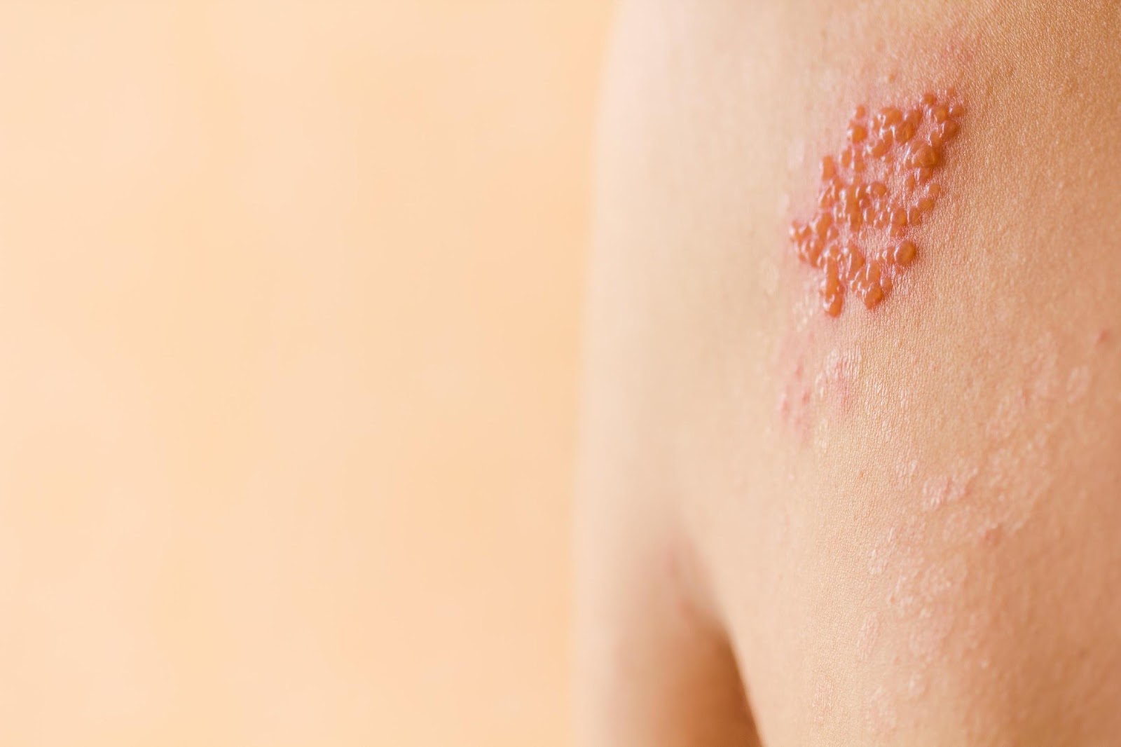
Shingles is a skin condition characterized by grouped vesicles with a reddish base in a band-like distribution usually affecting just one side of the body.
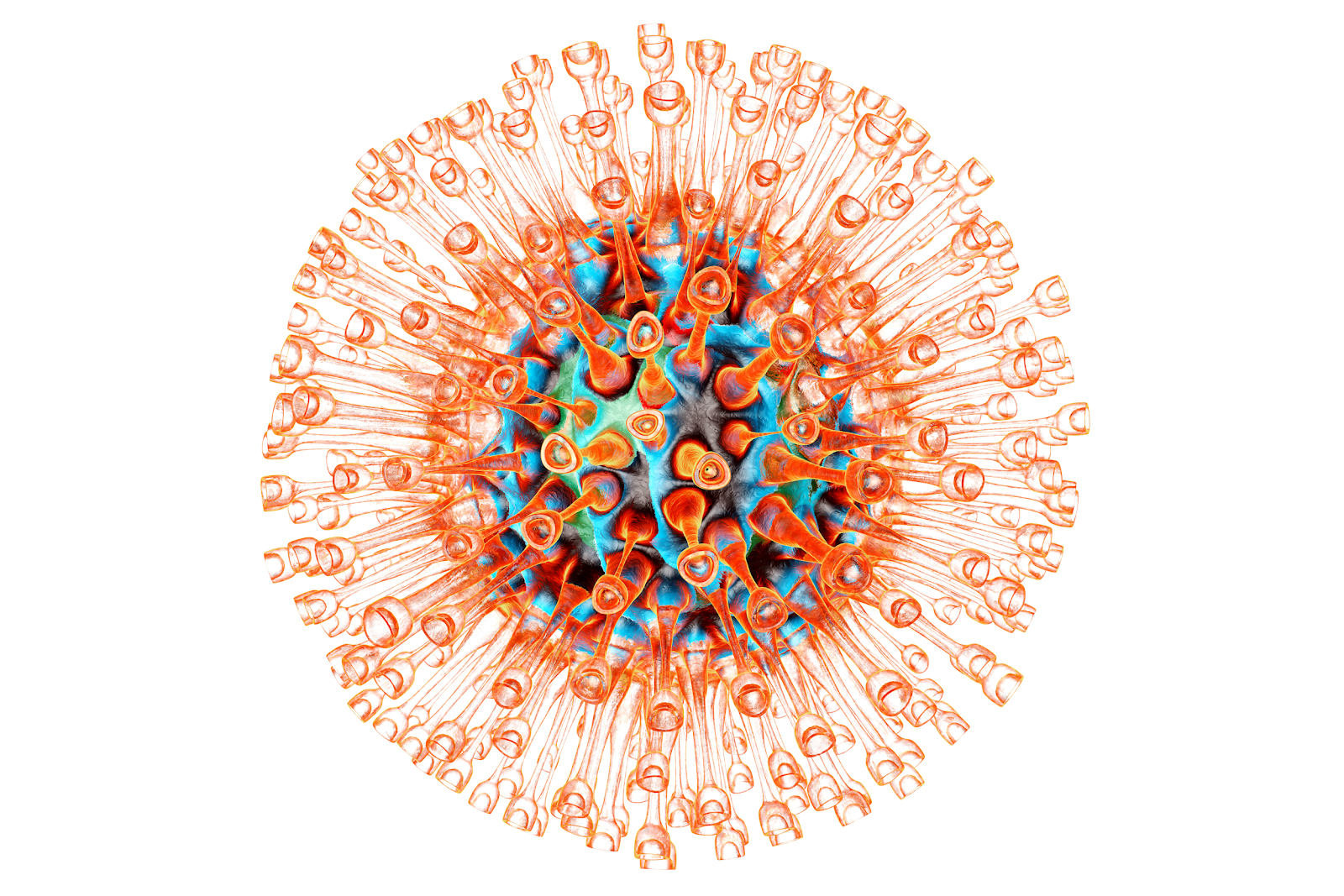
What causes Shingles?
Shingles is caused by Herpes Zoster Virus (HSV), which is also the virus that causes chickenpox. Shingles occur in people who already had chickenpox at one point in their lives. When a patient is healed from chickenpox clinically, HSV goes into a “resting” mode or latent mode in the neurons. When the same patient’s immune system experiences a decline, this virus can reactivate and can cause the expected skin lesions on the areas of the body supplied by the affected neurons.
Who gets Shingles?
Anyone can get Shingles but the following groups of people have increased risk in acquiring the disease
– elderly
– immunocompromised individuals (organ transplant patients, cancer patients)
– patients with immune-mediated conditions (systemic lupus erythematosus, rheumatoid arthritis etc.)
– patients undergoing chemotherapy, immunomodulators and corticosteroids
– HIV patients
What are the physical findings?
The lesions usually start as red patches on the affected body part in a unilateral distribution. After 12 to 24 hrs, grouped vesicles form on top of the patches. On the 3rd day, these vesicles can be filled with pus. In 7 to 10 days, brown crusts can form on top of the lesions. These crusts can persists for 2 to 3 weeks
What are the other signs and symptoms?
Is it infectious?
YES, Herpes Zoster Virus infection is infectious. A patient can transmit it to other people through direct contact with the vesicles until 7 days from the initial appearance of the skin lesions. It can also be airborne in some cases. The people who have exposure with patients with Shingles won’t develop Shingles, they will develop chickenpox.
It is important for patients to avoid exposure to pregnant mothers, elderly people, newborns and children.
Is it treatable?
YES, HZV infection is treatable.
– ORAL ANTI-VIRAL MEDICATIONS
It is important to seek consult with a dermatologist once the skin lesions are noted. Anti-viral medications are effective if given during the first 72 hours of the disease. It is proven that anti-viral therapy can decrease the duration and severity of the skin rash and associated pain. It also prevents the appearance of lesions to other parts of the body.
– TOPICAL TREATMENT OPTIONS
-can be beneficial during the acute phase to relieve pruritus and pain. It can also hasten the drying of
skin lesions

What are the possible complications?
Is there a vaccine available?
Zostavax ® (live attenuated Oka/Merck strain VZV zoster vaccine) is available and recommended to adults >60 y/o of age for prevention of herpes zoster and its complications, especially post-herpetic neuralgia.
Dr. Mara Padilla Evangelista-Huber, FPDS, FDSP, MClinRes
What gives color to our skin?
Melanin is the pigment molecule that gives our skin, hair and eyes color. It provides protection from ultraviolet radiation, which can cause premature skin aging (wrinkles, uneven skin) and skin cancer.
Melanin is made by cells in the skin called melanocytes, and one of the crucial steps involves an enzyme called tyrosinase. After melanin is made, it is transferred from melanocytes to the keratinocytes (skin cells).
What is hyperpigmentation?
Hyperpigmentation refers to excess pigmentation. This pertains to areas of skin that appear darker in color compared to other regions. It may develop anywhere in the body, may be small patches or be more widespread. While hyperpigmentation is not generally harmful, it can be a symptom of an underlying medical condition.
What causes hyperpigmentation?
There are many causes for hyperpigmentation. These triggers basically tell the melanocytes to make more melanin. One of the most common causative factors is sun exposure, which is why sun protection is very important.
Causes of hyperpigmentation:
Individuals with medium to darker-colored skin types are more prone to develop hyperpigmentation compared to those with lighter skin types because the former have more melanin to begin with.
What are the types of hyperpigmentation?
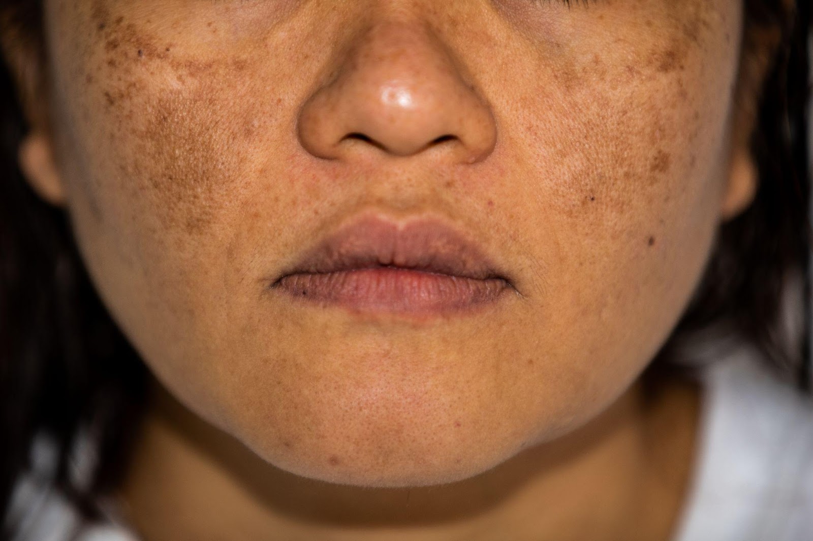
The most common types of hyperpigmentation are melasma, post-inflammatory hyperpigmentation, and sun spots or solar lentigines.
Melasma is one of the most common reasons for consultation with a dermatologist. The hyperpigmentation is usually on the face, specifically on the forehead, cheeks, nose, chin, but can occur elsewhere. Melasma mostly affects adult women and darker skin types. It can be caused by hormonal changes (e.g. pregnancy, use of oral contraceptives).
Post-inflammatory hyperpigmentation. This type of hyperpigmentation occurs after inflammation (common in acne and eczema patients) and injuries. You’ll see dark patches of skin on previously inflamed or reddish areas,
Sun spots or solar lentigines. This type of hyperpigmentation occurs in areas with high sun exposure like the face, forearms, hands. These are flat, round to oval dark patches, may be single or appear in a group.
How do we prevent and treat hyperpigmentation?
When dealing with hyperpigmentation, a two-way strategy is recommended: (1) to protect the skin and prevent hyperpigmentation from developing, and (2) to treat and correct existing hyperpigmentation.
Sunscreens, sun protection measures and topical anti-oxidants like vitamin C are very helpful in preventing hyperpigmentation. The cause of hyperpigmentation (e.g. acne, eczema, insect bites) must also be addressed.
When it comes to treating hyperpigmentation, a combination of these methods are beneficial: one is to inhibit tyrosinase (an important step in melanin synthesis), another is to inhibit the transfer of melanin from melanocytes to keratinocytes (skin cells), and lastly, to increase the renewal of skin so that new skin cells can replace the old, hyperpigmented skin cells.
Protect and prevent:
Treat: inhibit melanin transfer
Treat: tyrosinase inhibitor
Treat: increase cell renewal
What is hydroquinone?
Hydroquinone is a prescription-only drug considered a standard in the treatment of hyperpigmentation. While effective when used correctly, it may cause irritation, burning, stinging, and dryness – with higher concentrations posing higher risks. It is not recommended to use hydroquinone continuously due to the risk of ochronosis (paradoxical hyperpigmentation). The use of hydroquinone is only advised under the guidance of a dermatologist.
How long will it take for hyperpigmentation to resolve?
It depends. Hyperpigmentation may go away even without treatment within 3 to 24 months, but treatment can speed up the process. Hyperpigmentation may fully resolve or lighten considerably, but for others it may not completely disappear.
Hyperpigmentation: a final word
References:
Davis EC, Callender VD. Postinflammatory hyperpigmentation: a review of the epidemiology, clinical features, and treatment options in skin of color. J Clin Aesthet Dermatol. 2010;3(7):20-31.
Desai SR. Hyperpigmentation therapy: a review. J Clin Aesthet Dermatol. 2014;7(8):13-17.
Vashi NA, Wirya SA, Inyang M, Kundu RV. Am J Clin Dermatol. 2017 Apr; 18(2):215-230.
Huerth KA, Hassan S, Callender VD. Therapeutic Insights in Melasma and Hyperpigmentation Management. J Drugs Dermatol. 2019 Aug 1;18(8):718-729
Katrina Canlas-Estrella, MD, FPDS
Diet is frequently associated in various skin disorders. One way in which diet affects skin disorders is the concept of food allergy. Food allergy is defined by the US National Institute of Allergy and Infectious Diseases as: “an adverse health effect arising from an immune response that occurs reproducibly on exposure to a given food”. Simply put, this occurs when there is a breakdown of the body’s tolerance to food ingested1,2. Typical manifestations of a food allergy are skin reactions such as hives and itchiness, respiratory tract symptoms such as difficulty of breathing, and gastrointestinal tract symptoms such as vomiting and diarrhea. A severe and potentially fatal reaction called anaphylaxis, can also be experienced. While many advances have been made in understanding the mechanism, treatment and prevention of food allergy, the mainstay of treatment remains to be avoidance of the food allergen/s1-5.
Allergenic foods comprising more than 85% of food allergy are egg, milk, peanut, tree nuts (walnut, cashew, pistachio), fish, shellfish, sesame seed, soy, and wheat. Several studies identify egg allergy as the most prevalent4,5. In adults, allergies to certain fruits and vegetables are common3. Usually, many people outgrow their food allergies over time, such as hen egg and cow milk allergies. In contrast, peanut and tree nut allergies, along with shellfish allergy, are known to persist throughout life1-3. In addition, nickel is also a very common allergen and this could be found in certain foods such as oatmeal, beans, peas, soybeans, shellfish, and chocolate2,4,6.
A hypoallergenic diet is a diet composed of foods low in allergenicity. Specifically, it is free from soy, nuts, egg, dairy, corn, beef, gluten, shellfish, and citrus fruits. That said, patients on a strict hypoallergenic diet should be carefully monitored and properly managed so as to avoid nutritional deficiencies. In children, food allergies to milk, egg, soy, and wheat tend to disappear during late childhood and these specific foods may eventually be tolerated after 1 to 2 years. As mentioned, allergies to peanut, tree nuts, and shellfish typically persist and may be lifelong1-4.
There are certain skin diseases that can benefit from a hypoallergenic diet. Several studies have shown that certain food allergens can lead to an exacerbation of a patient’s dermatitis. Specifically, food allergy has been shown to be present in 20 to 80 percent of patients with atopic dermatitis (AD). Thus, a hypoallergenic diet may be helpful in patients with AD. Other skin diseases that may benefit from a hypoallergenic diet are systemic contact dermatitis (SCD) and allergic contact dermatitis (ACD). Furthermore, acute vesicular hand dermatitis may benefit from a diet low in nickel-rich foods2,4,6.
In order to find out if one has a food allergy and identify the specific food/s one is allergic to, one may consult with an allergologist. A detailed history and thorough physical examination are important, then skin prick testing (SPT) or radioallergosorbent test (RAST) can be done to identify potential food allergens. Once potential allergens have been identified, one must always be vigilant in checking food labels and ingredients in order to prevent the unfortunate manifestations of food allergy. In the event of an allergic reaction, rescue medication should always be available1-6.

References:
Kaimal, Sowmya, and Devinder Thappa. “Diet in Dermatology: Revisited.” Indian J Dermatol Venereol Leprol, vol. 76, no. 2, 2010, pp. 103–116.
by: Maria Elinor Grace Q. Sison, MD, FPDS
What is keratosis pilaris?
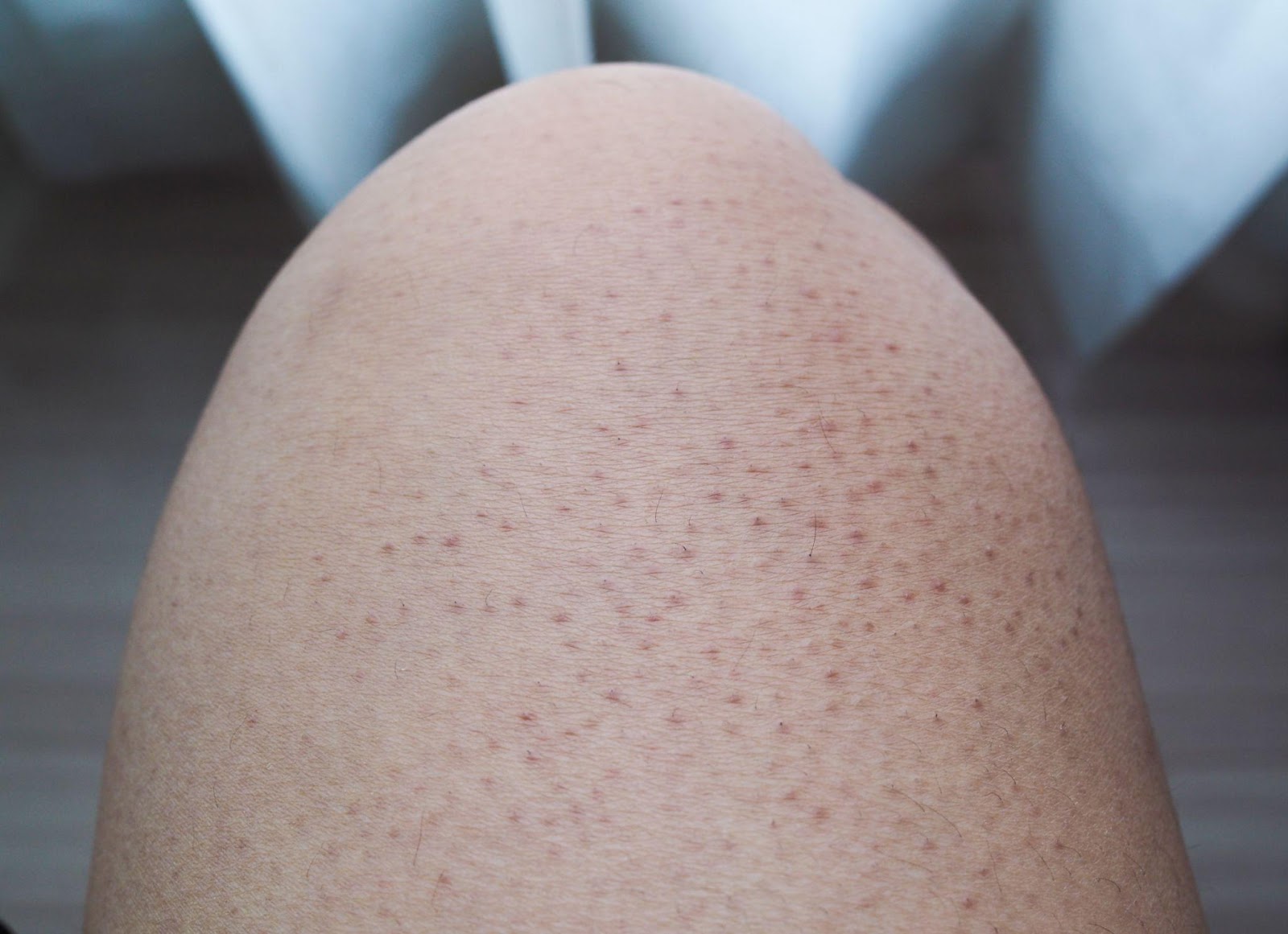
Keratosis pilaris or “chicken skin” is a common condition due to plugging of the follicles of our skin. It is common in children and can improve by late adolescence but is often persistent. It is strongly associated with several skin conditions such as ichthyosis vulgaris and atopic dermatitis. The cause of keratosis pilaris is not well understood.
How does keratosis pilaris present?
Keratosis pilaris presents with small bumps with varying degrees of redness. It affects lateral cheeks, extensor aspects of the upper arms, thighs, and buttocks. In children the face and arms are mainly involved while in adults the lesions are found in extensor arms and legs.
How is keratosis pilaris diagnosed?
Diagnosis is made through physical examination by the dermatologist based on the appearance of the lesions and their distribution.
What are the treatment options for keratosis pilaris?
Reference:Bruckner, AL. Keratosis Pilaris and Other Follicular Keratotic Disorders. In: Fitzpatrick’s Dermatology. 9th Ed. USA: McGraw-Hill.
By Dr. Coreen Copuyoc-Sampedro
Does wearing make-up ruin your skin? Back in the day, dermatologists were known to be against make-up or any form of cosmetic camouflage since many products then—think 80’s to early nineties —would cause a slew of unwanted effects such as acne, contact dermatitis, and hyperpigmentation. Instead, we would promote healthy natural looking skin sans any cosmetic product. However, the beauty and skincare market has grown exponentially and this preconception no longer applies. This not only means that the variety and number of products increased, but that many formulations have also gotten more sophisticated and safe. Going all-natural with just sunscreen on your face is definitely still a good way to go, but wearing make-up can actually work well with your skin if you know how to choose the right ones for your skin type. Aside from that, cosmetics actually help a lot of people as a confidence booster and as part of self-care. In fact, I am one such dermatologist that loves make-up almost (almost!) as much as skin care.
So how do you choose the right cosmetics for you? This is best figured out on a case to case basis but basically depends, first and foremost, on your skin type and the environment you’re in. If you have normal skin, almost any product may work well on you so just use the weather as your guide. Wearing oilier formulations may not work as well in a hot & humid environment such as in Manila for example, but these may be best for cold, breezy weather such as in Baguio. For those with oily skin, lightly formulated water-based foundations and medium-coverage powders may work best, provided that the skin is still hydrated. And for those with dry skin, oil-based formulas work best to camouflage anything but again, diligent hydration everyday is really the best way to ensure that make-up applies nicely. Combination skin is quite tricky because you will need to mix and match the products you use per area on your face. Creams or oil-based products applied on dry parts while lighter, more water-based products on the oily parts. Then lastly, the most difficult skin type to choose products for, is sensitive skin. The primary concern for sensitive skin would be avoidance of irritating or allergenic ingredients in make-up such as fragrances and parabens, while also choosing formulations that work well with your base skin type between oily, dry, or combination skin. I personally have sensitive, combination skin and have discovered that I like mousse or cream-gel formulations best, with light finishing powder on areas that are a bit oily.

At the end of the day, all of these are simply general guidelines and choosing your cosmetics will still depend on which ones are accessible to you and work well with your lifestyle and environment. Keep in mind though that the best foundation for make-up is still healthy, luminous skin. So I recommend allocating more time and budget for skin care and always remember to remove your make-up right when you get home. So, does wearing make-up ruin your skin? Well, the correct ones shouldn’t, and that’s the tea.